. 2007 Jan 26;8(1):3.
doi: 10.1186/1465-9921-8-3.

The role of pneumolysin in mediating lung damage in a lethal pneumococcal pneumonia murine model

Affiliations

The role of pneumolysin in mediating lung damage in a lethal pneumococcal pneumonia murine model

María Del Mar García-Suárez et al. Respir Res. .

Abstract

Background: Intranasal inoculation of Streptococcus pneumoniae D39 serotype 2 causes fatal pneumonia in mice. The cytotoxic and inflammatory properties of pneumolysin (PLY) have been implicated in the pathogenesis of pneumococcal pneumonia.

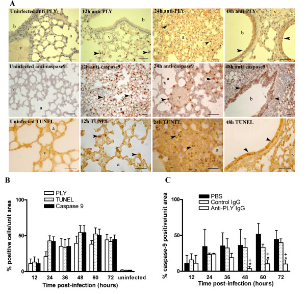
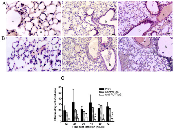
Methods: To examine the role of PLY in this experimental model we performed ELISA assays for PLY quantification. The distribution patterns of PLY and apoptosis were established by immunohistochemical detection of PLY, caspase-9 activity and TUNEL assay on tissue sections from mice lungs at various times, and the results were quantified with image analysis. Inflammatory and apoptotic cells were also quantified on lung tissue sections from antibody treated mice.

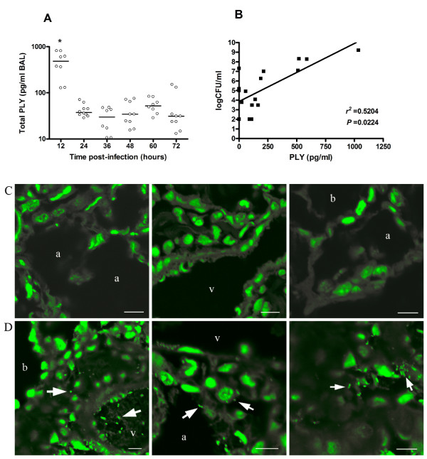
Results: In bronchoalveolar lavages (BAL), total PLY was found at sublytic concentrations which were located in alveolar macrophages and leukocytes. The bronchoalveolar epithelium was PLY-positive, while the vascular endothelium was not PLY reactive. The pattern and extension of cellular apoptosis was similar. Anti-PLY antibody treatment decreased the lung damage and the number of apoptotic and inflammatory cells in lung tissues.

Conclusion: The data strongly suggest that in vivo lung injury could be due to the pro-apoptotic and pro-inflammatory activity of PLY, rather than its cytotoxic activity. PLY at sublytic concentrations induces lethal inflammation in lung tissues and is involved in host cell apoptosis, whose effects are important to pathogen survival.


Figures

Figure 1
Figure 1
Concentrations of PLY and bacteria localization in lungs of mice infected with S. pneumoniae D39 serotype 2. (A) Amounts of total PLY were quantified in BAL after lysis of bacteria. Each symbol represents one mouse, and horizontal bars represent medians. Results are representative of three independent experiments. Groups were compared by nonparametric Mann-Whitney U test. * P < 0.05. (B) Correlation between CFU and PLY in BAL. Dots represent the means of CFU versus PLY concentration from three mice at the same time points of Fig. 1A Correlation was performed by nonparametric Spearman r-test. (C) Confocal images of lung tissue sections from uninfected mice. (D) Representative lung tissue sections from pneumococci infected mice showing intra-vessel, intra-cytoplasmic and intercellular bacteria localization. Blood vessel (v), alveolar space (a), bronchiole (b). Scale bars 8 μm. All images were captured after a Z-stack analysis of the samples.
Figure 2
Figure 2
Apoptosis in lung tissues of mice infected with S. pneumoniae D39 serotype 2. (A) Distribution patterns of PLY and apoptosis in representative lung sections from mice intranasally infected with S. pneumoniae D39 serotype 2. PLY was established by staining with anti-PLY rabbit antibodies. Apoptosis was assessed by active caspase-9 staining and in situ TUNEL assay. No staining was observed in lung tissues from uninfected mice. At 12 h post-infection, resident alveolar macrophages were positively stained with anti-PLY, anti-caspase-9, and TUNEL (arrow heads). Infiltrating leukocytes (24 h post-infection) and bronchial epithelium (48 h post-infection) were stained with anti-PLY, anti-caspase-9, and TUNEL, respectively (arrow heads). Note non-stained vascular endothelium. Blood vessel (v), alveolar space (a), bronchiole (b). Scale bars 50 μm. (B) Apoptosis and PLY in lung tissues from untreated mice during pneumococcal pneumonia. Apoptosis was identified by immunohistochemical detection of active caspase-9 and by in situ TUNEL assay. PLY was stained with anti-PLY rabbit antibodies. Adjacent sections were co-stained for co-localization of PLY, TUNEL, and caspase-9. Five sections were analyzed in each time point. Statistical differences were not found for a comparison of number of PLY, caspase-9, and TUNEL positive cells as determined by two-way ANOVA followed by the Bonferroni test. (C) Comparison of caspase-9 positive cells in lung tissues from anti-PLY IgG-, control IgG-, and PBS-treated mice. Percentage of caspase-9 stained cells was calculated with respect to total cells counted in random areas of lung tissue sections. Results are means ± SD of 3 mice and are representative of three independent experiments. *, P < 0.05 for a comparison of anti-PLY IgG-treated mice with PBS-treated mice, and +,P < 0.05 for a comparison of anti-PLY IgG with control IgG-treated mice, as determined by two-way ANOVA followed by the Bonferroni test.
Figure 3
Figure 3
Comparison of the level of inflammation of lung tissue among anti-PLY IgG-, control IgG-, and PBS-treated mice infected with S. pneumoniae D39 serotype 2. Histological appearance of representative lungs from mice infected intranasally with S. pneumoniae serotype 2 and treated with anti-PLY IgG (A), and control IgG (B). Numerous leukocytes can be seen in the peribronchial and perivascular areas, and considerable vascular distension and hemorrhage take place during the progression of pneumococcal colonization. Lungs of mice treated with anti-PLY IgG reveal alveoli, bronchioles, and vessels structurally normal, with no signs of acute inflammation and lower leukocyte infiltration. Blood vessel (v), alveolar space (a), bronchiole (b). Scale bars 50 μm. (C) Numbers of infiltrating cells were counted in random areas of lung tissue sections H&E-stained. Results are means ± SD of 3 mice and are representative of three independent experiments. *, P < 0.05 for a comparison of anti-PLY IgG- treated mice with PBS-treated mice, and +,P < 0.05 for a comparison of anti-PLY IgG with control IgG-treated mice, as determined by two-way ANOVA followed by the Bonferroni test.

References

    1. Cockeran R, Anderson R, Feldman C. Pneumolysin as a vaccine and drug target in the prevention and treatment of invasive pneumococcal disease. Arch Immunol Ther Exp. 2005;53:189–198. - PubMed
    1. Hirst RA, Kadioglu A, O'Callaghan C, Andrew PW. The role of pneumolysin in pneumococcal pneumonia and meningitis. Clin Exp Immunol. 2004;138:195–205. doi: 10.1111/j.1365-2249.2004.02611.x. - DOI - PMC - PubMed
    1. Palmer M. The family of thiol-activated, cholesterol-binding cytolysins. Toxicon. 2001;39:1681–1689. doi: 10.1016/S0041-0101(01)00155-6. - DOI - PubMed
    1. Rossjohn J, Gilbert RJC, Crane D, Morgan PJ, Mitchell TJ, Rowe AJ, Andrew PW, Paton JC, Tweten RK, Parker MW. The molecular mechanism of pneumolysin, a virulence factor from Streptococcus pneumoniae. J Mol Biol. 1998;284:449–461. doi: 10.1006/jmbi.1998.2167. - DOI - PubMed
    1. Balachandran P, Hollingshead SK, Paton JC, Briles DE. The autolytic enzyme LytA of Streptococcus pneumoniae is not responsible for releasing pneumolysin. J Bacteriol. 2001;183:3108–3116. doi: 10.1128/JB.183.10.3108-3116.2001. - DOI - PMC - PubMed

Publication types

LinkOut - more resources