. 2007;9(1):R12.
doi: 10.1186/bcr1645.

Hormone-induced protection of mammary tumorigenesis in genetically engineered mouse models

Affiliations

Hormone-induced protection of mammary tumorigenesis in genetically engineered mouse models

Lakshmanaswamy Rajkumar et al. Breast Cancer Res. 2007.

Abstract

Introduction: The experiments reported here address the question of whether a short-term hormone treatment can prevent mammary tumorigenesis in two different genetically engineered mouse models.

Methods: Two mouse models, the p53-null mammary epithelial transplant and the c-neu mouse, were exposed to estrogen and progesterone for 2 and 3 weeks, respectively, and followed for development of mammary tumors.

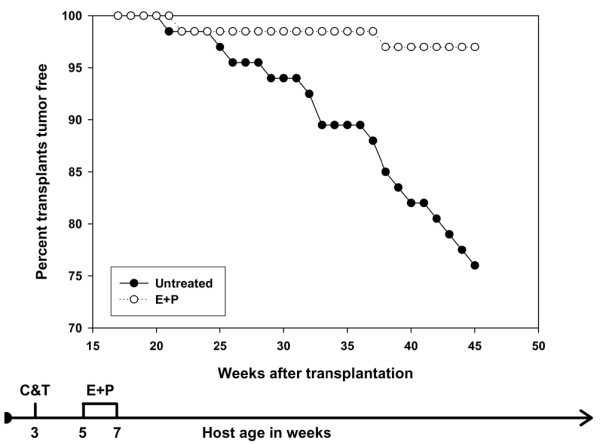
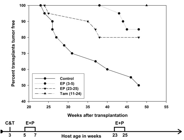
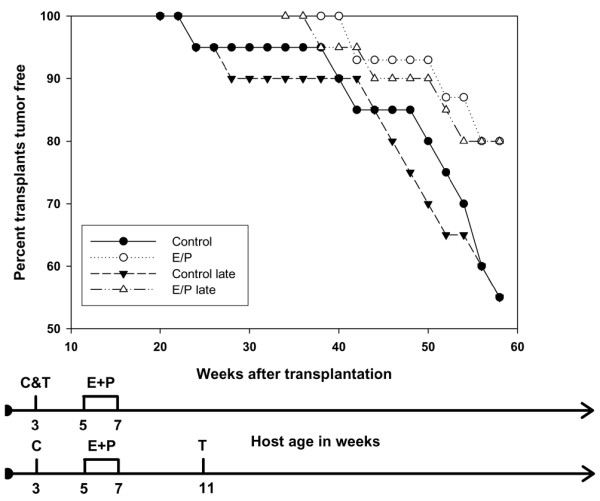
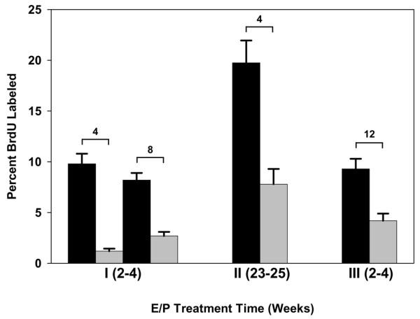
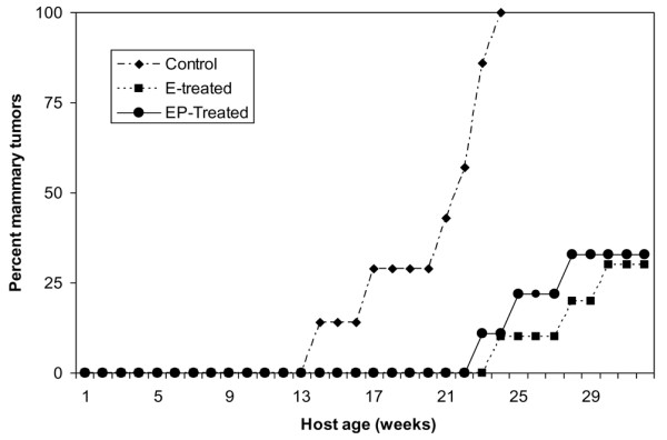
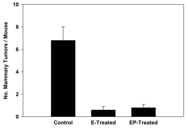
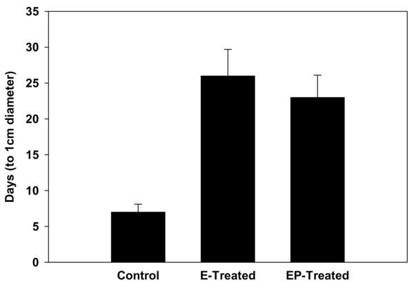
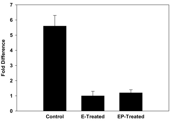
Results: In the p53-null mammary transplant model, a 2-week exposure to estrogen and progesterone during the immediate post-pubertal stage (2 to 4 weeks after transplantation) of mammary development decreased mammary tumorigenesis by 70 to 88%. At 45 weeks after transplantation, analysis of whole mounts of the mammary outgrowths demonstrated the presence of premalignant hyperplasias in both control and hormone-treated glands, indicating that the hormone treatment strongly affects the rate of premalignant progression. One possible mechanism for the decrease in mammary tumorigenesis may be an altered proliferation activity as the bromodeoxyuridine labeling index was decreased by 85% in the mammary glands of hormone-treated mice. The same short-term exposure administered to mature mice at a time of premalignant development also decreased mammary tumorigenesis by 60%. A role for stroma and/or systemic mediated changes induced by the short-term hormone (estrogen/progesterone) treatment was demonstrated by an experiment in which the p53-null mammary epithelial cells were transplanted into the cleared mammary fat pads of previously treated mice. In such mice, the tumor-producing capabilities of the mammary cells were also decreased by 60% compared with the same cells transplanted into unexposed mice. In the second set of experiments using the activated Her-2/neu transgenic mouse model, short-term estradiol or estradiol plus progesterone treatment decreased mammary tumor incidence by 67% and 63%, and tumor multiplicity by 91% and 88%, respectively. The growth rate of tumors arising in the hormone-treated activated Her-2/neu mice was significantly lower than tumors arising in non-hormone treated mice.

Conclusion: Because these experiments were performed in model systems that mimic many essential elements of human breast cancer, the results strengthen the rationale for translating this prevention strategy to humans at high risk for developing breast cancer.


Figures

Figure 1
Figure 1
Effect of short-term hormone treatment on tumorigenesis in p53-null mammary epithelial transplants. The data are the sum of two identical experiments (experiments 1 and 1A). Mice treated with estradiol and progesterone had a significant decrease in tumor incidence (p < 0.05). Filled circles, untreated; open circles, hormone-treated. The flow diagram illustrates the experimental treatment plan. C, clear mammary gland; T, transplant; E, estrogen; P, progesterone.
Figure 2
Figure 2
Effect of short-term hormone treatment on gland morphology in p53-null transplants. (a,b) Mammary whole mounts were prepared 4 weeks after the removal of hormones in mice from experiment 1A. The ductal organization at the histological and microscopic (hematoxylin/eosin-stained sections, not shown) was similar in the untreated (a) and treated (b) mice (original magnification ×2). (c–f) Whole mounts were prepared at 45 weeks after transplantation in transplants that had not yet developed palpable tumors. A large number of transplants in the untreated group (46%) (original magnifications ×1 (c) and ×2 (d)) and in the treated group (42%) (original magnifications ×1 (e) and ×2 (f)) contained areas of ductal hyperplasias (arrows).
Figure 3
Figure 3
The effect of duration of hormone exposure on tumorigenesis in p53-null mammary epithelial transplants. The effect of hormone exposure was compared with that of a hormone receptor antagonist, tamoxifen. Mice treated with estradiol and progesterone, either early or late, or treated with tamoxifen, had a significant decrease in tumor incidence (p < 0.05). Filled circles and solid line, control; filled circles and dotted line, early hormone exposure; inverted triangles and broken line, late hormone exposure; upright triangle, tamoxifen. The flow diagram illustrates the experimental treatment plan.
Figure 4
Figure 4
The effect of hormone-induced systemic changes on tumorigenesis in p53-null mammary epithelial transplants. Mammary epithelial transplants in a host exposed to estradiol and progesterone before transplantation (open triangles and dot-dashed line) also had a significant decrease in tumor incidence (p < 0.05) compared with mice not exposed to the hormones (filled triangles and broken line). The flow diagrams illustrate the experimental treatment plans.
Figure 5
Figure 5
Bromodeoxyuridine-labeling index (number labeled per 500 cells) in hormone-treated p53-null mammary epithelial transplants. Transplants from each experiment (1, 2 and 3) were assayed for bromodeoxyuridine. The black bars represent untreated transplants, the gray bars hormone-treated transplants. The number above each pair of bars indicates the number of weeks after the removal of hormones. In experiment 3, 12 weeks after the removal of hormone represents 8 weeks after transplantation. There were four transplants per treatment group. Five hundred cells were counted in each transplant. All four comparisons were significantly different (p < 0.05).
Figure 6
Figure 6
Immunohistochemistry of bromodeoxyuridine-labeled mammary epithelial cells. The brown nuclei (arrows) represent the uptake of bromodeoxyuridine into cells undergoing DNA synthesis. (a) Untreated; (b) treated with estrogen plus progesterone.
Figure 7
Figure 7
Effect of short-term hormone treatment on mammary carcinogenesis in the activated Her-2/neu transgenic mice. Mice treated with estradiol alone (7/21) or estradiol plus progesterone (9/24) had a significant decrease in the incidence of mammary tumors compared with the controls (20/20) at 8 to 9 months of age (p < 0.01).
Figure 8
Figure 8
Effect of short-term hormone treatment on mammary cancer multiplicity in the activated Her-2/neu transgenic mice. The group of mice (n = 21) treated with estradiol alone developed a total of 13 mammary tumors and the group of mice (n = 24) treated with estradiol plus progesterone developed a total of 20 mammary tumors by 8 to 9 months of age; in comparison, the group of control mice (n = 20) developed a total of 136 mammary tumors (p < 0.005). Results are expressed as means ± SEM.
Figure 9
Figure 9
Effect of short-term hormone treatment on tumor growth rate in the activated Her-2/neu transgenic mice. Treatment with estradiol alone or estradiol plus progesterone significantly increased the time taken for the mammary tumors to grow from initial palpation to a diameter of 1 cm in comparison with the controls. Results are expressed as means ± SEM.
Figure 10
Figure 10
Cyclin D1 mRNA expression levels in mammary tumors arising in activated Her-2/neu mice. There was an approximately sixfold increase in the level of cyclin D1 in mammary tumors arising in control mice in comparison with tumors arising in hormone-treated mice. Results are expressed as means ± SEM.

Comment in

Similar articles

Cited by

References

    1. Beaston GL. On the treatment of inoperable cases of carcinoma of the mamma: suggestions for a new method of treatment with illustrative cases. Lancet. 1896;ii:104–107. - PMC - PubMed
    1. Howell A, Cuzick J, Baum M, Buzdar A, Dowsett M, Forbes JF, Hoctin-Boes G, Houghton J, Locker GY, Tobias JS, ATAC Trialists' Group Results of the ATAC (Arimidex, Tamoxifen, Alone or in Combination) trial after completion of 5 years' adjuvant treatment for breast cancer. Lancet. 2005;365:60–62. doi: 10.1016/S0140-6736(05)74803-0. - DOI - PubMed
    1. Willett WC, Rockhill B, Hawkinson SE, Hunter D, Colditz GA. Nongenetic factors in the causation of breast cancer. In: Harris JA, Lippman ME, Morrow M, Osborne CK, editor. Diseases of the Breast. 3. Philadelphia: Lippincott, Williams & Wilkins; 2004. pp. 223–276.
    1. Huggins C, Moon RC, Morii S. Extinction of experimental mammary cancer. I. Estradiol-17β and progesterone. Proc Natl Acad Sci USA. 1962;48:379–386. doi: 10.1073/pnas.48.3.379. - DOI - PMC - PubMed
    1. Sivaraman L, Medina D. Hormone-induced protection against breast cancer. J Mammary Gland Biol Neoplasia. 2002;7:77–92. doi: 10.1023/A:1015774524076. - DOI - PubMed

Publication types