. 2007;9(1):R13.
doi: 10.1186/bcr1646.

Telomeric DNA induces apoptosis and senescence of human breast carcinoma cells

Affiliations

Telomeric DNA induces apoptosis and senescence of human breast carcinoma cells

Mina Yaar et al. Breast Cancer Res. 2007.

Abstract

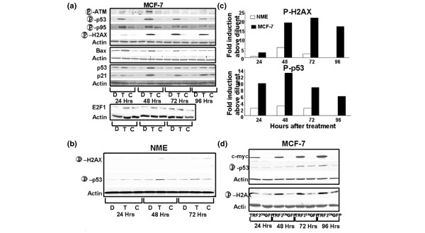
Introduction: Cancer is a leading cause of death in Americans. We have identified an inducible cancer avoidance mechanism in cells that reduces mutation rate, reduces and delays carcinogenesis after carcinogen exposure, and induces apoptosis and/or senescence of already transformed cells by simultaneously activating multiple overlapping and redundant DNA damage response pathways.

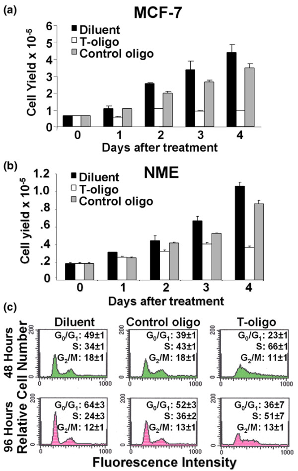
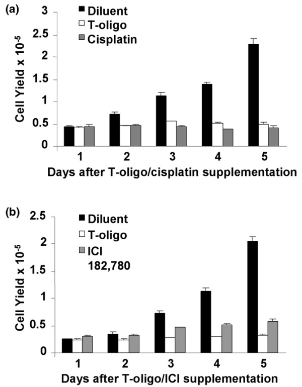
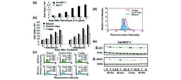
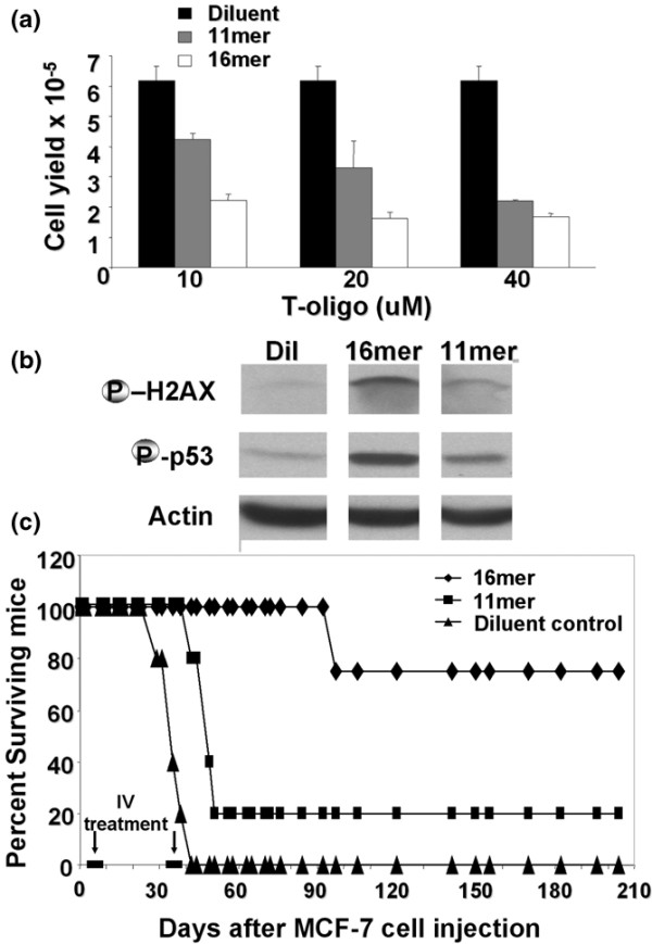
Methods: The human breast carcinoma cell line MCF-7, the adriamycin-resistant MCF-7 (Adr/MCF-7) cell line, as well as normal human mammary epithelial (NME) cells were treated with DNA oligonucleotides homologous to the telomere 3' overhang (T-oligos). SCID mice received intravenous injections of MCF-7 cells followed by intravenous administration of T-oligos.

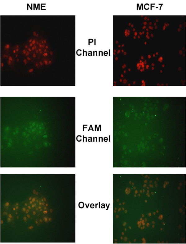
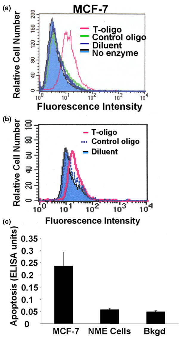
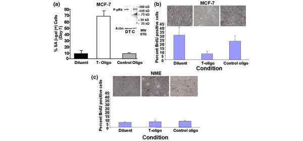
Results: Acting through ataxia telangiectasia mutated (ATM) and its downstream effectors, T-oligos induced apoptosis and senescence of MCF-7 cells but not NME cells, in which these signaling pathways were induced to a far lesser extent. In MCF-7 cells, experimental telomere loop disruption caused identical responses, consistent with the hypothesis that T-oligos act by mimicking telomere overhang exposure. In vivo, T-oligos greatly prolonged survival of SCID mice following intravenous injection of human breast carcinoma cells.

Conclusion: By inducing DNA damage-like responses in MCF-7 cells, T-oligos provide insight into innate cancer avoidance mechanisms and may offer a novel approach to treatment of breast cancer and other malignancies.


Figures

Figure 1
Figure 1
T-oligos localize to nuclei. MCF-7 and normal mammary epithelial (NME) cells were supplemented with fluorescein phosphoramidite (FAM) labeled GTTAGGGTTAG and uptake was determined 30 to 60 minutes after supplementation. Multicolor fluorescent microscopy showed that FAM-labeled T-oligos accumulated in the nucleus as determined by colocalization of propidium iodide (PI) nuclear staining (red) with FAM (green), producing an orange color. As expected, no green fluorescence was observed in control, diluent-treated cells.
Figure 2
Figure 2
T-oligo decreases cell yield and induces S phase arrest in MCF-7 cells. Preconfluent cultures of (a) MCF-7 cells and (b) normal mammary epithelial (NME) cells were treated with 40 μM of GTTAGGGTTAG (T-oligo), CTAACCCTAAC (control oligo) or diluent alone and cell yields were determined at different intervals after oligonucleotide addition. Compared to controls, within 4 days T-oligo decreased cell yields 87 ± 9% (MCF-7; p < 0.04, n = 4) and 65.5 ± 0.01% (NME; p < 0.03, n = 2). (c) MCF-7 cells were treated as above and cells were collected for up to 96 hours for FACScan analysis after a single treatment at time 0. Averages and standard deviations were determined from duplicate samples. T-oligo increased the percent of cells in the S phase of the cycle as early as 48 hours after stimulation (p < 0.001, n = 2) and for at least 96 hours after stimulation (p < 0.04, n = 2).
Figure 3
Figure 3
T-oligo induces apoptosis of MCF-7 cells. MCF-7 and normal mammary epithelial (NME) cells were treated as above. Apoptosis was determined by TUNEL analysis and by the level of cytoplasmic DNA-histone complexes 96 hours after a single treatment at time 0. (a) MCF-7 cells underwent apoptosis as observed by the prominent shift in fluorescent peak. (b) In contrast, there was only a minimal shift in the fluorescent peak of NME cells. (c) There is a significant increase in the levels of cytoplasmic DNA-histone complexes in T-oligo-treated MCF-7 cells compared to diluent-treated MCF-7 cells (p < 0.02, n = 3). In contrast, DNA-histone complexes in NME cells were not significantly increased above the background (Bkgd) level (p = 0.89).
Figure 4
Figure 4
T-oligo induces senescence of MCF-7 cells. (a,b) MCF-7 and (c) normal mammary epithelial (NME) cells were treated with T-oligo once for seven days and then were either fixed and stained for senescence-associated (SA) β-galactosidase (β-gal) activity (a), or were supplemented with fresh medium lacking oligonucleotides and 5-bromo-2'-deoxyuridine (BrdU) incorporation (b,c) and retinoblastoma protein phosphorylation (inset) were determined. (a) In MCF-7 cells, T-oligo induced senescence of 61.3 ± 7.7% cells, compared to diluent and control oligo in which 6.3 ± 3.8% and 12 ± 2% of the cells, respectively, were senescent, as determined by SA β-gal activity (p < 0.015). (b) In MCF-7 cells, after T-oligo removal and provision of medium without T-oligo, only 7.4 ± 3.3% of cells displayed BrdU incorporation compared to 30.4 ± 9.8% and 23 ± 6.4% of diluent and control oligo pretreated cells, respectively (p < 0.05). (c) In NME cells, BrdU incorporation did not differ between T-oligo and diluent-treated cultures (7.1 ± 2.2% versus 6.3 ± 1.1% and 7.8 ± 1.2%, respectively; p = 0.5). Inset: MCF-7 cells were treated as above. Despite supplementation of fresh medium lacking T-oligo, no phosphorylation of retinoblastoma protein (pRb) was detected in T-oligo pretreated cultures. C, control-oligo; D, diluent; T, T-oligo.
Figure 5
Figure 5
T-oligo and telomeric repeat binding factor dominant negative construct (TRF2DN) activate cell cycle- and apoptosis-regulatory proteins in breast cancer cells. (a) Compared with control oligo (C) and diluent (D) alone, T-oligo (T) substantially induced the phosphorylation of p53 (Ser15), p95/Nbs1 (Ser343) and H2AX (Ser139) and the level of E2F1. In MCF-7 cells, T-oligo also induced the phosphorylation of ataxia telangiectasia mutated (ATM; Ser1981) and the levels of p53 and the p53-dependent p21 and BAX proteins. (b) Compared with control oligo (C) and diluent (D), T-oligo (T) only slightly induced the phosphorylation of p53 and H2AX in normal mammary epithelial (NME) cells. (c) Graphic representation of band intensity as fold induction above diluent. (d) adenovirus (Ad)TRF2DN, but not the control Ad-green fluorescent protein (GFP) vector, induced the phosphorylation of H2AX and p53 in MCF-7 cells. c-myc expression confirms successful transfection of the cells with AdTRF2DN. Actin serves as a loading control. One of three representative experiments is shown.
Figure 6
Figure 6
T-oligo effect on MCF-7 cells is comparable to that of cisplatin and ICI 182,780. MCF-7 cells were treated with cisplatin (10 μM), the anti-estrogen ICI 182,780 (100 nM) or T-oligo (40 μM) once and cell yields were determined at different intervals. (a) Within 5 days cisplatin decreased MCF-7 yield by 82 ± 0.1% and T-oligo did so by 79 ± 0.02% compared to diluent. The difference between the T-oligo and cisplatin effects was not significant (p = 1.0). (b) Compared to diluent, ICI 182,780 or T-oligo caused decreases in cell yield of MCF-7 cells of 72 ± 0.1% and 84 ± 0.02%, respectively. T-oligo was significantly more effective than ICI 182,780 (p < 0.005).
Figure 7
Figure 7
T-oligo affects adriamycin resistant breast cancer cells. (a) Unlike MCF-7 cells, Adr/MCF-7 cells proliferate in the presence of adriamycin. (b) Supplementation with T-oligo on day 0 decreased Adr/MCF-7 cell yields in both the presence and absence of adriamycin for at least 96 hours (58% ± 14%, p < 0.03, n = 4). (b) T-oligo also increased the percent of cells in the S phase within 48 hours and through 96 hours (p < 0.04, n = 2) and (d) induced apoptosis as determined by TUNEL analysis at 96 hours. (e) T-oligo induced the phosphorylation of p53 and H2AX comparably to MCF-7 cells (Figure 5a). C, control-oligo; D, diluent; T, T-oligo.
Figure 8
Figure 8
T-oligo increases survival of mice injected intravenously with MCF-7 cells. (a) MCF-7 cells received fresh serum-containing medium supplemented with increasing concentrations (10 to 40 μM) of the 11mer or 16mer T-oligos or diluent alone and were harvested at 96 hours. At 10 μM and 20 μM, the 16mer inhibited MCF-7 yield substantially more than the 11mer, and 20 μM 16mer reduced cell yields comparably to 40 μM 11mer. (b) MCF-7 cells were supplemented with 11mer or 16mer T-oligos (20 μM) or diluent (Dil) alone. Within 48 hours both T-oligos induced H2AX and p53 phosphorylation, but this occurred to a greater extent with the 16-base T-oligo. (c) SCID mice inoculated intravenously with MCF-7 cells received intravenous T-oligo or diluent injections as per the Materials and methods. Doses giving comparable growth inhibition at 96 hours were used: 20 μM 16mer versus 40 μM 11mer, equivalent to 60 versus 120 nmoles injected into the estimated murine 1.5 ml peripheral blood volume. Both T-oligos increased survival of the mice, with the 16mer having a greater effect.

Similar articles

Cited by

References

    1. Jemal A, Murray T, Ward E, Samuels A, Tiwari RC, Ghafoor A, Feuer EJ, Thun MJ. Cancer statistics, 2005. CA Cancer J Clin. 2005;55:10–30. - PubMed
    1. Seidman AD. Systemic treatment of breast cancer. Two decades of progress. Oncology (Williston Park) 2006;20:983–990. discussion 991–982, 997–988. - PubMed
    1. Calvo FA, Meirino RM, Orecchia R. intraoperative radiation therapy part 2. Clinical results. Crit Rev Oncol Hematol. 2006;59:116–127. - PubMed
    1. Weber DC, Ares C, Lomax AJ, Kurtz JM. Radiation therapy planning with photons and protons for early and advanced breast cancer: an overview. Radiat Oncol. 2006;1:22. doi: 10.1186/1748-717X-1-22. - DOI - PMC - PubMed
    1. Hickey BE, Francis D, Lehman MH. Sequencing of chemotherapy and radiation therapy for early breast cancer. Cochrane Database Syst Rev. 2006:CD005212. - PubMed

Substances