. 2007 Apr;1768(4):772-93.
doi: 10.1016/j.bbamem.2006.12.002. Epub 2006 Dec 15.

Expansion of signal transduction by G proteins. The second 15 years or so: from 3 to 16 alpha subunits plus betagamma dimers

Affiliations

Expansion of signal transduction by G proteins. The second 15 years or so: from 3 to 16 alpha subunits plus betagamma dimers

Lutz Birnbaumer. Biochim Biophys Acta. 2007 Apr.

Abstract

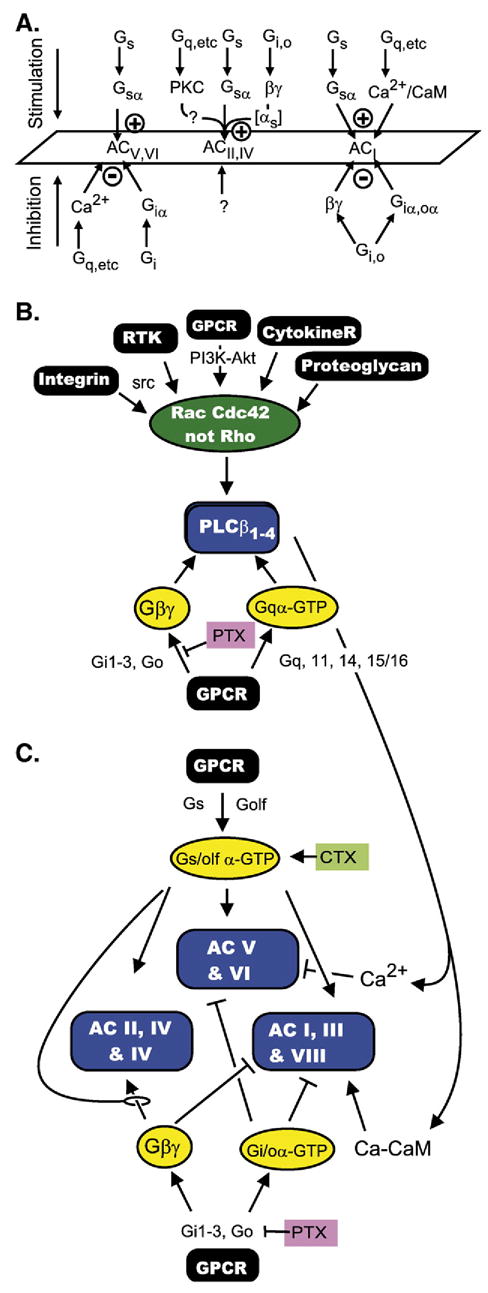
The first 15 years, or so, brought the realization that there existed a G protein coupled signal transduction mechanism by which hormone receptors regulate adenylyl cyclases and the light receptor rhodopsin activates visual phosphodiesterase. Three G proteins, Gs, Gi and transducin (T) had been characterized as alphabetagamma heterotrimers, and Gsalpha-GTP and Talpha-GTP had been identified as the sigaling arms of Gs and T. These discoveries were made using classical biochemical approaches, and culminated in the purification of these G proteins. The second 15 years, or so, are the subject of the present review. This time coincided with the advent of powerful recombinant DNA techniques. Combined with the classical approaches, the field expanded the repertoire of G proteins from 3 to 16, discovered the superfamily of seven transmembrane G protein coupled receptors (GPCRs) -- which is not addressed in this article -- and uncovered an amazing repertoire of effector functions regulated not only by alphaGTP complexes but also by betagamma dimers. Emphasis is placed in presenting how the field developed with the hope of conveying why many of the new findings were made.


Figures

Fig. 1
Fig. 1
Signal transduction of hormone binding to receptor into activation of adenylyl cyclase by the Gs G protein as understood in 1983. Gs is shown to undergo a GTP–GDP driven subunit dissociation–reassociation cycle, in which the role of the receptor is to keep the system cycling by promoting the Mg2+-induced exchange of GTP for GDP and in which the role of GTPα is both the activation of the enzyme and its own time-limited inactivation that comes about when it hydrolyzes GTP to GDP. (Adapted from Hildebrandt et al., 1984a [119]).
Fig. 2
Fig. 2
(A) Mg2+ mimics the effect of receptor to accelerate the activation of Gs by GMP-P(NH)P (from Iyengar and Birnbaumer, 1981 [9]). (B) Effect of the hormone–receptor complex to reduce the concentration at which Mg2+ facilitates activation of Gs by GMP-P(NH)P. Liver membranes were treated with N-ethyl maleimide to inactivate its adenylyl cyclase and then subjected to a 10-min incubation with GMP-P(NH)P in the absence and presence of glucagon at increasing concentrations of Mg2+ shown on the figure. Gs in the incubated membranes was extracted and analyzed for activity in a cyc reconstitution assay. Note the hormone induced left shift in the Mg2+ concentration curve. Inset, expansion of lower range of the Mg2+ concentration axis (from Iyengar and Birnbaumer, 1982 [10]). (C) Mg2+ increases the rate at which GMP-P(NH)P activates purified Gs. Purified human erythrocyte Gs was preincubated for varying times with GMP-P(NH)P at varying concentrations of Mg2+. Progressive activation was then quantified in diluted aliquots using the cyc reconstitution assay. Note that the rate of activation increased as Mg2+ concentration was increased. Top, data are shown with time as the x-axis variable. Bottom, the data are shown with Mg2+ concentration as the x-axis variable (adapted from Codina et al., 1984 [11]).
Fig. 3
Fig. 3
Heterotrimeric G protein α subunits are discovered to be structural relatives of other regulatory GTPases. Tα, transducin α; Gα37, Goα: Sc ras1, ras1 of the baker’s yeast Saccharomyces cerevisiae; Ypt1, the Saccharomyces cerevisiae homologue of rab1 (rat brain ras like protein 1) (Adapted from Hurley et al., 1984a [16]).
Fig. 4
Fig. 4
The rates of phosphoinositide mobilization and diacylglycerol formation are consistent with a primary effect of receptor on phospholipase C to promote phosphorylase activation. (Adapted from Thomas et al., 1983 [50]).
Fig. 5
Fig. 5
Inositol 1,4,5-tris phosphate (IP3) promotes rapid release of Ca2+ from the stores of permeabilized pancreatic acinar cells. Permeabilized cells were incubated in minimum medium containing physiologic concentration of Ca2+. At the indicated time, ATP was added as an energy source for the cells’ intracellular Ca2+ uptake system, causing ambient Ca2+ to fall. Addition of IP3 caused release of Ca2+ from internal stores — which equilibrated with extracellular Ca2+. As IP3 was hydrolyzed by phosphatases, Ca2+ was re-accumulated in stores. Ca2+ was monitored with a Ca2+ electrode. Traces in the bottom panel show that carbachol, an agonist of the pancreatic muscarinic receptor, mobilizes Ca2+ from the same pool affected by the externally added IP3. Other experiments had previously shown that pancreatic muscarinic receptors cause phosphoinositide hydrolysis and mobilize Ca2+ from internal stores (Adapted from Streb et al., 1983 [52]).
Fig. 6
Fig. 6
Receptors that activate the Gs-adenylyl cyclase signaling pathway share with receptors that activate the G protein-PLC signaling pathway the property of changing their affinity for agonist upon addition of guanine nucleotides. (A) GTP and Mg regulation of the β-adrenergic receptor (βAR) in membranes with (I) and without (II) the Gs G protein. The figure depicts the competitive inhibition by the βAR agonist (−)-isoproterenol of the binding of the βAR probe 125I-hydroxybenzylpindolol (125I-HYP) to βARs of wild type (I) and cyc (II) S49 cell membranes. The incubations were carried out in the absence and presence of 5 mM MgCl2, 10 μM GTP and the combination of MgCl2 and GTP. Mg2+ and GTP do not affect the affinity of IHYP for the βAR (not shown). Note: Mg2+ increased the affinity of the βAR for the competing agonist, and GTP, which by itself had no effect, interfered with the effect of Mg2+. This phenomenon is mimicked by non-hydrolizable analogs of GTP and by GDP, and is commonly referred to as the “GTP-shift in agonist affinity”. There are receptors, such as the glucagon receptor (Rojas and Birnbaumer, 1985 [182]), that show high agonist affinity also in the absence of Mg2+ but shift to low agonist affinity when GTP is added. The high affinity state of the βAR is induced by its interaction with the Gs G protein; the shift is absent in cells lacking Gs (AII) (adapted from Hildebrandt et al., 1984b [118]; Hildebrandt et al., 1984a [119]). (B) The non-hydrolyzable GTP analogue GMP-P(NH)P causes a GTP affinity shift in the liver α1-adrenergic receptor known to trigger activation of phosphoinositide hydrolysis and promote Ca2+ mobilization, suggesting the existence of a G protein responsible for phosphoinositide and Ca2+ mobilization (adapted from Good-hardt et al., 1980 [58]).
Fig. 7
Fig. 7
Bradykinin, an agonist that promotes phosphonositide hydrolysis and Ca2+ mobilization, activates a low Km GTPase in membranes from NG108-15 cells. (A and B) Hydrolysis of [γ-32P]GTP in the absence and presence of 1 μM bradykinin as a function of GTP concentration expressed as % [γ-32P]GTP hydrolyzed (A) or pmol of GTP hydrolyzed (B). (C) [γ-32P]GTP hydrolyzed in the presence of 0.3 μM unlabelled GTP as a function of varying the concentrations of the agonist bradykinin or the inactive des-Arg9-bradykinin analogue. (Adapted from Grandt et al., 1986 [53]).
Fig. 8
Fig. 8
(A) Musings that may have happened during the “making” of the Gq/11 family of G proteins into signal transducers of PLCβ-activating receptors. (B) The Gq/11 signal transduction pathway.
Fig. 9
Fig. 9
Linear diagram of a consensus open reading frame (yellow box) encoding α subunits of the family of heterotrimeric signal transducing G proteins. Cyan box, sequences of ras for which homologous sequences are present in Gα subunits. id, identity box sequence; its composition is expanded below; the id sequence is identical for αs, αi1-αi3, αt1, αt2, α-olf, αo. Deviant amino acids in αq, 11, 14, 12, 13, and z are shown. Black boxes, regions involved in guanine nucleotide binding. R*, location of the Arg ADP-ribosylated by cholera toxin in αs, α-olf and transducin α. The seven C-terminal amino acids of α subunits are expanded to include the Cys at position-4 which is ADP-ribosylated by pertussis toxin (C*). Several mutations that have been informative for the understanding of the G protein mediated signal transduction process are highlighted: G to V in the GAGES motif reduces GTPase activity; R* to C or H reduces GTPase activity, as does ADP-ribosylation, which allow for receptor independent activation by GTP; Q to L in the DVGGQ motif suppresses GTPase activity and confers transforming activity to mutant α subunits; G to T in the DVGGQ motif confers dominant negative properties; G to A in the DVGGQ motif (H21a mutant in αs, also reverse UNC) impedes activation by GTP or non-hydrolyzable GTPγS, but does not interfere with the G protein mediated GTP shift in receptor-agonist affinity; R to P at position-6 from the C terminus, UNC mutation, uncouples αs form receptor without interfering with activation by non-hydrolyzable GTP analogues. N to I and D to N in the FLNKXD motif change nucleotide association–dissociation dynamics. Mutants of Q in the DVGGQ motif, G in the GAGES motif and R* confer transforming activity in transfection assays and are referred to as gsp and gip when they reside in the αs and αi subunits, respectively.
Fig. 10
Fig. 10
(A) Signaling by mating factor receptors Ste2p and Ste3p, activated by α and a factor, respectively, is transduced in haploid Saccharomyces cerevisiae cells by an αβγ signal transducing G protein encoded in genes GPA1, STE4 and STE18. α or a factor binding to their respective receptors leads to growth arrest. In this G protein coupled system the signaling arm is the βγ dimer (Step:Ste18p (F)) and not the Gpa1p.GTP (αGTP) complex (Jiang et al., 1993 [80]). If mating does not occur, cells resume growth, a phenomenon referred to as adaptation. The SSTE gene product, a GTPase activating protein for Gpa1p, participates in this adaptation process. Ste18p(F), denotes that Ste18p, the product of the STE18 gene, is farnesylated. (B) Mutant loss of function stte2Δ cells are supersensitive to mating factor, seen in the formation of large halos of growth arrested cells. Supersensitivity can be suppressed by expression of mammalian RGS proteins (adapted from Druey et al., 1996 [93]).
Fig. 11
Fig. 11
(A) Activation and de-activation of muscarinic inwardly rectifying K+ channels is accelerated by the GTPase activating protein RGS4. The effect of a pulse of acetylcholine on the development of K+ currents was measured by the two electrode voltage clamp method. Top trace, K+ currents evoked by addition of acetylcholine (ACh) during the time-span shown, were recorded in an oocyte that that had been injected with cRNAs encoding the M2 muscarinic receptor and the Kir3.1 and Kir3.2 K+ channel subunits. Bottom trace, K+ currents evoked by acetylcholine in an oocyte that had been injected in addition with cRNA encoding mammalian RGS4. Note faster response to ACh addition and faster deactivation upon ACh wash-out. (Adapted from Doupnik et al., 1997 [98]). (B) Simulation of HR complex formation [p=f(t)] from H and R as a function of time using the formulas and rate constants shown. (C) Same as (B) but calculated for koff values spanning from 0.01 to 30 s−1. Note that at increasing koff equilibrium is reached faster and also, that the p(eq) values decrease. (D) Same as (C) but normalizing by expressing p(t) as a function of its value at equilibrium.
Fig. 12
Fig. 12
Stimulation of LARG’s guanine nucleotide exchange activity by G12α depends on Tec kinase, but that of G13α does not. Top: linear diagram of RhoGEF proiesn with relative location of the DHPH GEF domains, the RGSL domain (for extended RGS domain) and, when present, the PDZ domain. Bottom, in vitro stimulation of GDP release from RhoA by recombinant G12α and G13α in the absence and presence of Tec kinase and ATP. Tec was prepared by expressing a myc-tagged Tec cDNA in COS cells and concentrating the expressed protein by immunoprecipitation. G12α, G13α and LARG (without its N-terminal PDZ domain) were purified from Sf9 insect cells infected with the corresponding recombinant baculovirus. For further details see (Suzuki et al., 2003 [111]).
Fig. 13
Fig. 13
The G12/13 signaling pathway. Rho-GEF activities involve their DHPH domains. DH, Dbl-domain; PH, PH domain; BM, Btk domain; Y, Tyrpsine; pY, phosphotyrosine. G12α and Gβγ are shown as activators of the Tec kinases Tec and Btk. The interaction has been shown to involve the PHBM domain of the Tec kinase (Tsukada et al., 1994 [114]; Langhans-Rajasekaran et al., 1995 [183]; Jiang et al., 1998 [113]).
Fig. 14
Fig. 14
Stimulation of PLC activity by G protein βγ dimers. (A) Either GTPγS or transducin βγ (Gtβγ) stimulate phosphinositidase activity in a high speed cytosol supernatant from differentiated HL60 cells. Formation of inositol 1,4,5-trisphosphate (InsP3) correlates with hydrolysis of labeled phosphatidyl-4,5-bisphosphate (PtdInsP2) added to the incubations. (B) Dose–response relation for the stimulatory effect of transducin βγ. (C) The effect of transducin βγ is prevented by increasing concentrations of GDP-liganded transducin α (Gtα-GDP). (Adapted from Camps et al., 1992 [147]).
Fig. 15
Fig. 15
Stimulation of recombinant PLCβ activity by the Rac2 and Cdc42Hs members of the Rho family of GTPases. (A) Diagram of domain distribution along the PLCβ polypeptide chain. PH, plekstrin homology domain; 4EF, four EF hand folds, X and Y catalytic domains; C2, Ca2+-binding C2 domain. (B) Recombinant PLCβ2Δ, lacking the C-terminal portion that confers responsiveness to Gq/11α subunits, was incubated with substrate and recombinant Rac2: :LyGDI dimers in the presence of GDP, GDP+ recombinant β5γ2 dimers, GTPγS, or GTPγS+ recombinant β5γ2 dimers. (C) Recombinant PLCB2Δ was incubated with substrate and increasing concentrations of either recombinant β1γ2, or recombinant Cdc42Hs. Recombinant proteins were synthesized in SF9 cells infected with the corresponding recombinant baculoviruses, extracted, and purified to homogeneity by conventional techniques. For details see Illenberger et al., 2003 [149]. (Adapted from Figs. 9A, 8B and 7B) in Illenberger et al., 2003 [149]).
Fig. 16
Fig. 16
(A) Adenylyl cyclases are targets of multiple regulatory signaling pathways and respond differently, depending on which group they belong to. The figure shows these differences as summarized in 1994 (adapted from Taussig et al., 1994 [154]. (B) The β-type phosphoinositide-specific phospholipases Cβ are targets of three different signaling pathways (for G protein triggered signals see text; for signals impinging on the Rac-Cdc42 GTPases see Cerione (2004 [184]) and references therein), the Gqα family of α subunits and Gβγ dimers. (C) Example of cross talk from the Gq (Gqα), Gi (Gβγ) and Cdc42-Rac signalling pathways to the Gs signaling pathway may generate increases of decreases in cAMP levels depending on the subset of adenylyl cyclases expressed in the target cell.

Similar articles

Cited by

References

    1. Birnbaumer L. The Discovery of Signal Transduction by G Proteins. A personal account and an overview of the initial findings and contributions that led to our present understanding. Biochim Biophys Acta Biomembranes. 2007;1768:756–771. doi: 10.1016/j.bbamem. 2006.09.027. (in press) - DOI - PMC - PubMed
    1. Ovchinnikov YA. Rhodopsin and bacteriorhodopsin: Strucnture-Function Relationships. FEBS Letters. 1982;148:179–191. - PubMed
    1. Dixon RAF, Kobilka BK, Strader DJ, Benovic JL, Dohlman HC, Frielle T, Bolanowski MA, Bennett CD, Rands E, Diehl RE, Mumford RA, Slater EE, Sigal IS, Caron MG, Lefkowitz RJ, Strader CD. Cloning of the Gene and cDNA for Mammalian Beta-adrenergic Receptor and Homology with Rhodopsin. Nature. 1986;321:75–79. (March 11; published, May issue). - PubMed
    1. Yardeen Y, Rodriguez H, Wong SKF, Brandt DR, May DC, Burnier J, Harkins RN, Chen EY, Ramachandran J, Ullrich A, Ross EM. The Avian beta-Adrenergic Receptor: Primary Structure and Topology. Proc Natl Acad Sci USA. 1986;83:6795–6799. Acceptd June 9, published Sept Issue. - PMC - PubMed
    1. Maguire ME, Van Arsdale PM, Gilman AG. An Agonist-Specific Effect of Guanine Nucleotides on Binding to the Beta Adrenergic Receptor. Mol Pharmacol. 1976;12:335–339. - PubMed

Publication types

MeSH terms

Substances

LinkOut - more resources