. 2007 Apr;26(1):134-45.
doi: 10.1016/j.nbd.2006.12.008. Epub 2006 Dec 20.

BACE1 gene deletion prevents neuron loss and memory deficits in 5XFAD APP/PS1 transgenic mice

Affiliations

BACE1 gene deletion prevents neuron loss and memory deficits in 5XFAD APP/PS1 transgenic mice

Masuo Ohno et al. Neurobiol Dis. 2007 Apr.

Abstract

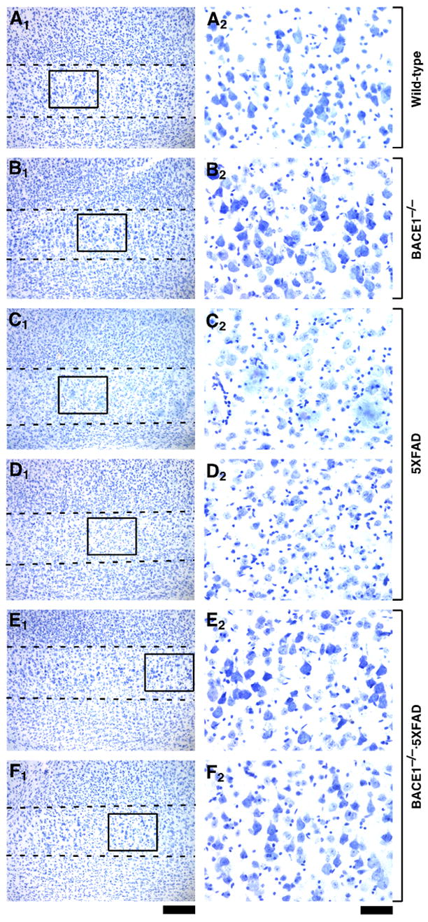
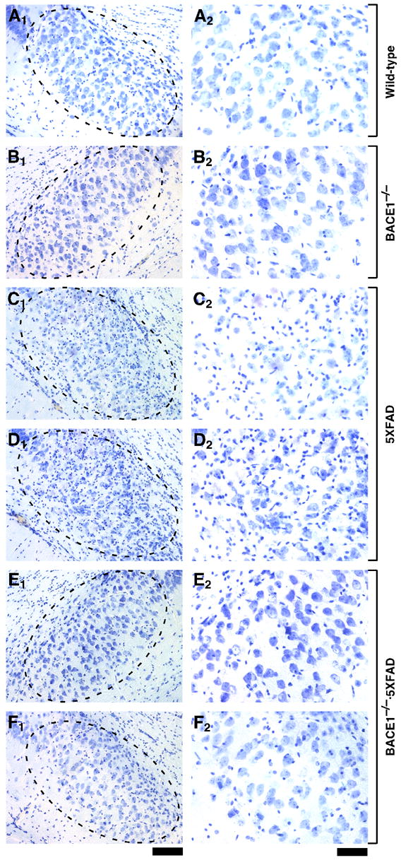
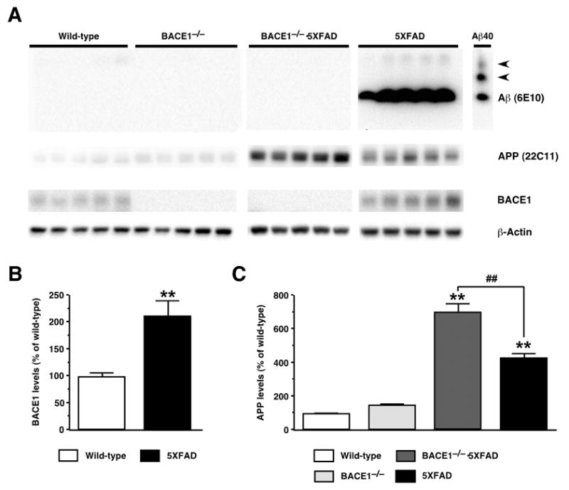
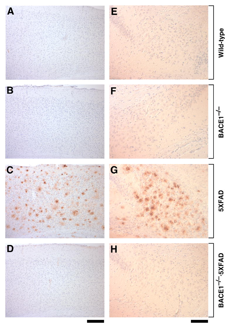
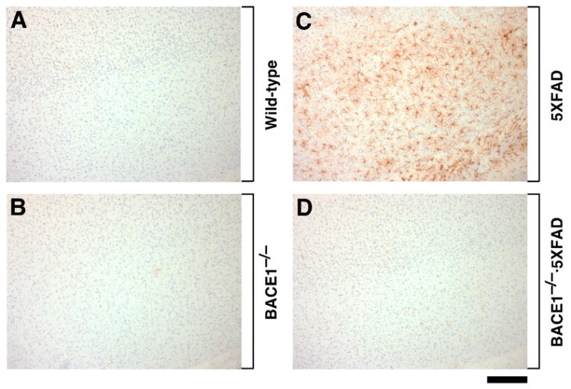
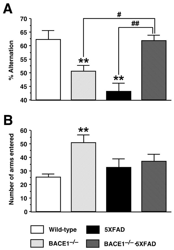
Evidence suggests that beta-amyloid (Abeta) peptide triggers a pathogenic cascade leading to neuronal loss in Alzheimer's disease (AD). However, the causal link between Abeta and neuron death in vivo remains unclear since most animal models fail to recapitulate the dramatic cell loss observed in AD. We have recently developed transgenic mice that overexpress human APP and PS1 with five familial AD mutations (5XFAD mice) and exhibit robust neuron death. Here, we demonstrate that genetic deletion of the beta-secretase (BACE1) not only abrogates Abeta generation and blocks amyloid deposition but also prevents neuron loss found in the cerebral cortex and subiculum, brain regions manifesting the most severe amyloidosis in 5XFAD mice. Importantly, BACE1 gene deletion also rescues memory deficits in 5XFAD mice. Our findings provide strong evidence that Abeta ultimately is responsible for neuron death in AD and validate the therapeutic potential of BACE1-inhibiting approaches for the treatment of AD.


Figures

Fig. 1
Fig. 1
BACE1 null mutation prevents neuronal loss in the cerebral cortex of 5XFAD transgenic mice. Sagittal brain sections from wild-type control (A), BACE1−/− (B), 5XFAD (C,D), and BACE1−/−·5XFAD (E,F) mice at 18 months of age were stained with cresyl violet. Two different mice are presented for each of the 5XFAD and BACE1−/−·5XFAD genotypes. Shown are photomicrographs of cortical layer 5 (A1–F1: areas between the dashed lines) and higher magnification of pyramidal neurons in areas identified by respective rectangles (A2–F2). Note that large pyramidal neurons are nearly completely lost in layer 5 of 5XFAD mice as compared with the other three genotypes. In contrast, large pyramidal neurons are rescued to wild-type levels in BACE1−/−·5XFAD brains. Scale bar = 200 μm in A1–F1; 50 μm in A2–F2.
Fig. 2
Fig. 2
BACE1 null mutation prevents neuronal loss in the subiculum of 5XFAD transgenic mice. Sagittal brain sections from wild-type control (A), BACE1−/− (B), 5XFAD (C,D), and BACE1−/−·5XFAD (E,F) mice at 18 months of age (the same brains shown in Fig. 1) were stained with cresyl violet. Two different mice are presented for each of the 5XFAD and BACE1−/−·5XFAD genotypes. Shown are photomicrographs of the subiculum (A1–F1: areas within dashed ovals) and respective higher magnification images (A2–F2). Note that large pyramidal neurons are significantly reduced in number in subiculum of 5XFAD mice as compared with the other three genotypes; numbers of glia are increased, as indicated by the high density of small cresyl violet-stained nuclei. In contrast, the quantity of large pyramidal neurons in subiculum of BACE1−/−·5XFAD brains appears similar to that of wild-type brains, demonstrating rescue of neuron loss by BACE1 gene deletion. Scale bar = 100 μm in A1–F1; 50 μm in A2–F2.
Fig. 3
Fig. 3
Effects of BACE1 null mutation on cerebral Aβ, BACE1 and full-length APP levels in 5XFAD transgenic mice. (A) Immunoblot analyses of protein extracts (15 μg/lane) from hemibrain homogenates of wild-type control, 5XFAD, BACE1−/−·5XFAD, and BACE1−/− mice at 18 months of age. Shown are immunoblots for Aβ (top panels; anti-total Aβ antibody 6E10), APP (second from top panels; anti-mouse and human APP antibody 22C11), BACE1 (third from top panels; anti-BACE1 antibody BACE1-Cat), and actin (bottom panels; anti-actin antibody AC-15). 5XFAD brain homogenates show a strong Aβ band, while Aβ is below the level of detection by immunoblot analysis in brain extracts from the other three genotypes. The lane labeled “Aβ40” contains 100 ng of synthetic Aβ40 for use as a positive control; the two slower migrating bands (arrowheads) likely represent multimeric Aβ assemblies. (B) Quantification of BACE1 blots in which band intensities were measured by phosphorimaging and expressed as percentage of wild-type control levels. Note that BACE1 levels are significantly elevated in 5XFAD brains. (C) Quantification of APP blots in which band intensities were measured by phosphorimaging and expressed as percentage of wild-type control levels. Note that while 5XFAD mice overexpress human APP approximately fourfold relative to endogenous mouse APP protein, levels of APP are elevated even further in BACE1−/−·5XFAD mice. Data are presented as the mean ± SEM of 5 animals. **P < 0.01 (vs. wild-type controls), ##P < 0.01 (vs. BACE1−/−·5XFAD).
Fig. 4
Fig. 4
BACE1 null mutation prevents amyloid deposition in 5XFAD transgenic mice. Sagittal brain sections from wild-type control (A,E), BACE1−/− (B,F), 5XFAD (C,G) and BACE1−/−·5XFAD (D,H) mice at 18 months of age were immunostained with antibody specifically recognizing the Aβ42 C-terminus and counterstained with hematoxylin. Shown are photomicrographs of the layers of the cerebral cortex (A–D) and subiculum (E–H). Note that 5XFAD sections have robust Aβ42 staining in both brain regions, while BACE1−/−·5XFAD brain is protected from Aβ42 accumulation. Scale bar = 200 μm in A–D; 100 μm in E–H.
Fig. 5
Fig. 5
BACE1 null mutation prevents gliosis in 5XFAD transgenic mice. Sagittal sections of cerebral cortex from wild-type control (A), BACE1−/− (B), 5XFAD (C) and BACE1−/−·5XFAD (D) mice at 18 months of age were immunostained for astrocyte marker GFAP and counterstained with hematoxylin. Note that 5XFAD cortex shows extensive astrogliosis, while BACE1−/−·5XFAD brain has control levels of astrocytes. Scale bar = 200 μm.
Fig. 6
Fig. 6
BACE1 null mutation prevents memory deficits in 5XFAD transgenic mice. (A) Spatial working memory of mice at 15–18 months of age was tested by spontaneous alternation performance in the Y-maze. BACE1−/−·5XFAD mice are rescued completely back to wild-type levels of alternation in the Y-maze. Note also that both BACE1−/− mice and 5XFAD mice show significantly lower levels of alternation performance as compared to wild-type control and BACE1−/−·5XFAD mice, although the deficit for 5XFAD tends to be more severe. (B) Total number of arm entries indicates that BACE1−/− mice show significantly higher levels of exploratory activity in the Y-maze as compared to wild-type control mice. Data are presented as the mean ± SEM of 7–14 animals. **P < 0.01 (vs. wild-type controls), #P < 0.05, ##P < 0.01 (vs. BACE1−/−·5XFAD).

Similar articles

Cited by

References

    1. Akiyama H, Barger S, Barnum S, Bradt B, Bauer J, Cole GM, Cooper NR, Eikelenboom P, Emmerling M, Fiebich BL, Finch CE, Frautschy S, Griffin WS, Hampel H, Hull M, Landreth G, Lue L, Mrak R, Mackenzie IR, McGeer PL, O’Banion MK, Pachter J, Pasinetti G, Plata-Salaman C, Rogers J, Rydel R, Shen Y, Streit W, Strohmeyer R, Tooyoma I, Van Muiswinkel FL, Veerhuis R, Walker D, Webster S, Wegrzyniak B, Wenk G, Wyss-Coray T. Inflammation and Alzheimer’s disease. Neurobiol Aging. 2000;21:383–421. - PMC - PubMed
    1. Apostolova LG, Dutton RA, Dinov ID, Hayashi KM, Toga AW, Cummings JL, Thompson PM. Conversion of mild cognitive impairment to Alzheimer disease predicted by hippocampal atrophy maps. Arch Neurol. 2006;63:693–699. - PubMed
    1. Ashe KH. Learning and memory in transgenic mice modeling Alzheimer’s disease. Learn Mem. 2001;8:301–308. - PubMed
    1. Berger-Sweeney J, McPhie DL, Arters JA, Greenan J, Oster-Granite ML, Neve RL. Impairments in learning and memory accompanied by neurodegeneration in mice transgenic for the carboxyl-terminus of the amyloid precursor protein. Brain Res Mol Brain Res. 1999;66:150–162. - PubMed
    1. Bobinski M, Wegiel J, Tarnawski M, Bobinski M, Reisberg B, de Leon MJ, Miller DC, Wisniewski HM. Relationships between regional neuronal loss and neurofibrillary changes in the hippocampal formation and duration and severity of Alzheimer disease. J Neuropathol Exp Neurol. 1997;56:414–420. - PubMed

Publication types

MeSH terms

Substances