. 2007 Apr 1;35(2):467-77.
doi: 10.1016/j.neuroimage.2006.12.010. Epub 2006 Dec 16.

Diffusion tensor imaging of post mortem multiple sclerosis brain

Affiliations

Diffusion tensor imaging of post mortem multiple sclerosis brain

Klaus Schmierer et al. Neuroimage. .

Abstract

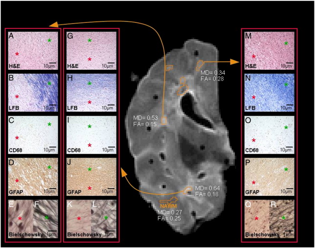
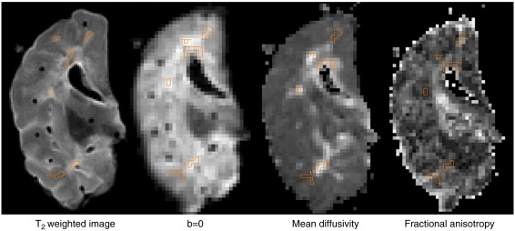
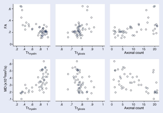
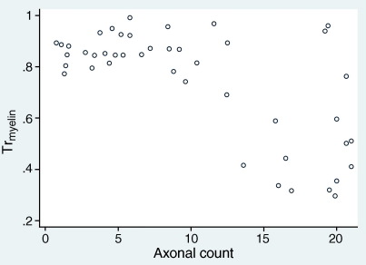
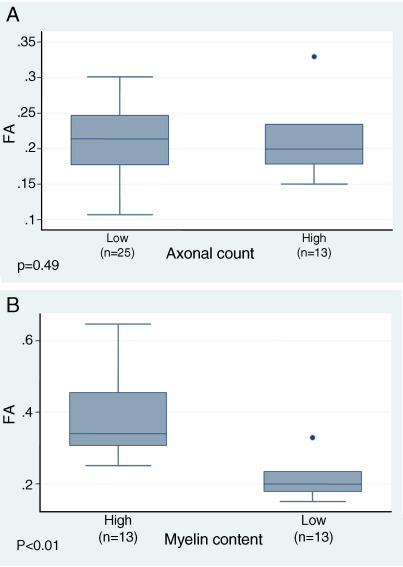
Magnetic resonance imaging (MRI) is being used to probe the central nervous system (CNS) of patients with multiple sclerosis (MS), a chronic demyelinating disease. Conventional T(2)-weighted MRI (cMRI) largely fails to predict the degree of patients' disability. This shortcoming may be due to poor specificity of cMRI for clinically relevant pathology. Diffusion tensor imaging (DTI) has shown promise to be more specific for MS pathology. In this study we investigated the association between histological indices of myelin content, axonal count and gliosis, and two measures of DTI (mean diffusivity [MD] and fractional anisotropy [FA]), in unfixed post mortem MS brain using a 1.5-T MR system. Both MD and FA were significantly lower in post mortem MS brain compared to published data acquired in vivo. However, the differences of MD and FA described in vivo between white matter lesions (WMLs) and normal-appearing white matter (NAWM) were retained in this study of post mortem brain: average MD in WMLs was 0.35x10(-3) mm(2)/s (SD, 0.09) versus 0.22 (0.04) in NAWM; FA was 0.22 (0.06) in WMLs versus 0.38 (0.13) in NAWM. Correlations were detected between myelin content (Tr(myelin)) and (i) FA (r=-0.79, p<0.001), (ii) MD (r=0.68, p<0.001), and (iii) axonal count (r=-0.81, p<0.001). Multiple regression suggested that these correlations largely explain the apparent association of axonal count with (i) FA (r=0.70, p<0.001) and (ii) MD (r=-0.66, p<0.001). In conclusion, this study suggests that FA and MD are affected by myelin content and - to a lesser degree - axonal count in post mortem MS brain.


Figures

Fig. 1
Fig. 1
Correlation of MRI and histopathology in post mortem multiple sclerosis brain (same case as in Fig. 2). On the T2-weighted scan of a coronal brain slice seven exemplary regions of interest (marked in orange) were identified as either normal-appearing white matter (NAWM) or white matter lesions (WMLs). Three WMLs are matched to respective histopathological sections, which were stained for hematoxylin and eosin (H&E), Luxol fast blue (LFB), CD68, glial fibrillary acid protein (GFAP) and Bielschowsky silver impregnation. Sections A–F illustrate a demyelinated WML with moderate infiltration by CD68-positive cells indicating chronic inflammatory activity (chronic active WML), and an axonal loss of 74% (compared to NAWM). Sections G–L show a hypo-cellular demyelinated lesion with very little inflammatory activity (chronic inactive WML). Axonal loss in this WML was 93%. Sections M–R show a remyelinated WML again with very little inflammatory activity (remyelinated WML) and an axonal loss of only 42%. Due to their high magnification (× 1250) images of Bielschowsky stained sections were divided into two halves (WML on the left and NAWM on the right). All other sections cover WMLs (red asterisks) as well as NAWM (green asterisks). MD = mean diffusivity × 10− 3 [mm2/s]; FA = fractional anisotropy.
Fig. 2
Fig. 2
T2-weighted (T2w) MRI, b = 0, mean diffusivity (MD) and fractional anisotropy (FA) maps of post mortem multiple sclerosis brain. On T2w MRI of a coronal brain slice, seven exemplary regions of interest (ROI; marked in orange) were identified as either normal-appearing white matter (NAWM) or white matter lesions (WMLs). ROIs were visually matched with respective ROI on b = 0 images, and then co-registered to the MD and FA maps. One of the five WMLs seen on T2w MRI (asterisk) could not be detected on diffusion maps.
Fig. 3
Fig. 3
Correlation between diffusion indices and quantitative histology in post mortem multiple sclerosis brain (white matter lesions, normal-appearing white matter). The plots illustrate the association of fractional anisotropy (FA) and mean diffusivity (MD) with (i) transmittance of sections stained for Luxol fast blue (Trmyelin, inversely proportional to myelin content), (ii) transmittance of sections immuno-stained for glial fibrillary acidic protein (Trgliosis, inversely proportional to severity of gliosis) and (iii) axonal count. See Table 3 for correlation coefficients.
Fig. 4
Fig. 4
Correlation between transmittance (Tr) of sections stained for Luxol fast blue (Trmyelin, inversely proportional to myelin content) and axonal count in post mortem multiple sclerosis brain (white matter lesions, normal-appearing white matter).
Fig. 5
Fig. 5
Box-plots of fractional anisotropy (FA) in regions of interest (ROIs) with (A) low myelin content (i.e., high transmittance of sections stained for Luxol fast blue, Trmyelin) and (B) high axonal count. ROIs with low myelin content and high axonal count are those below and above the respective means over all ROIs. ROIs with low myelin content are shown divided into those with low and high axonal counts: no difference in FA is evident between these subgroups (p = 0.49). ROIs with high axonal counts are shown divided into those with high and low myelin content: ROIs with high myelin content have a higher FA than ROIs with low myelin content (p < 0.01).

References

    1. Baltagi H. John Wiley and Sons; New York: 1995. Econometric Analysis of Panel Data.
    1. Bammer R., Skare S., Newbould R., Liu C., Thijs V., Ropele S., Clayton D.B., Krueger G., Moseley M.E., Glover G.H. Foundations of advanced magnetic resonance imaging. NeuroRx. 2005;2:167–196. - PMC - PubMed
    1. Barkhof F., Brück W., de Groot C.J., Bergers E., Hülshof S., Geurts J.J.G., Polman C.H., van der Valk P. Remyelinated lesions in multiple sclerosis: magnetic resonance image appearance. Arch. Neurol. 2003;60:1073–1081. - PubMed
    1. Barnes D., Munro P.M., Youl B.D., Prineas J.W., McDonald W.I. The longstanding MS lesion. A quantitative MRI and electron microscopic study. Brain. 1991;114:1271–1280. - PubMed
    1. Basser P.J., Mattiello J., LeBihan D. Estimation of the effective self-diffusion tensor from the NMR spin echo. J. Magn. Reson., Ser. B. 1994;103:247–254. - PubMed

Publication types