

Post-translational modification regulates prostaglandin D2 synthase apoptotic activity: characterization by site-directed mutagenesis
- PMID: 17259069
- PMCID: PMC1805777
- DOI: 10.1016/j.prostaglandins.2006.09.006
Post-translational modification regulates prostaglandin D2 synthase apoptotic activity: characterization by site-directed mutagenesis
Abstract
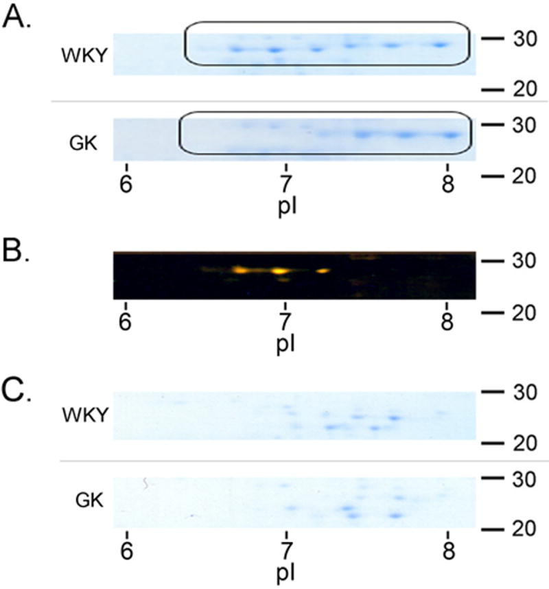
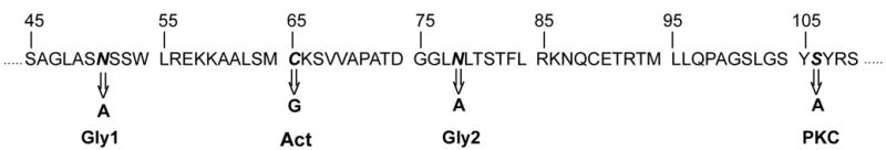
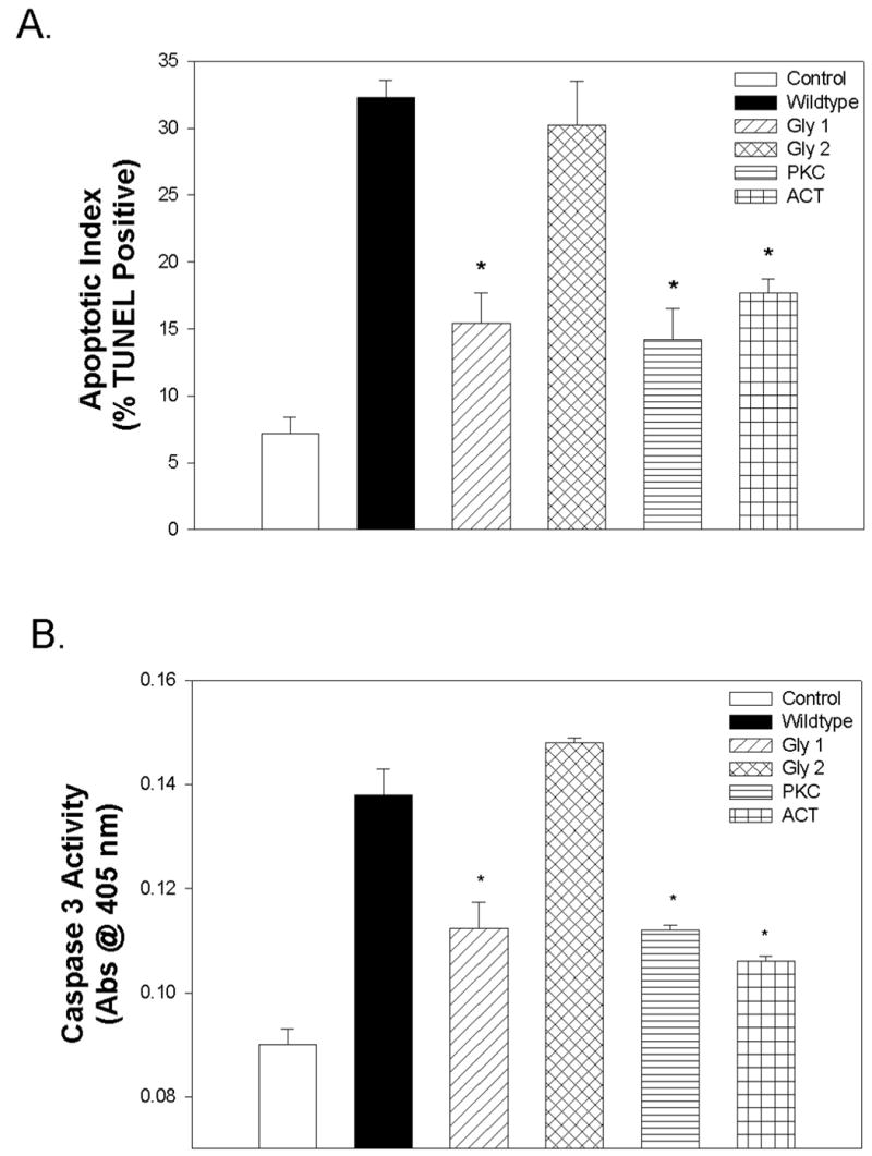
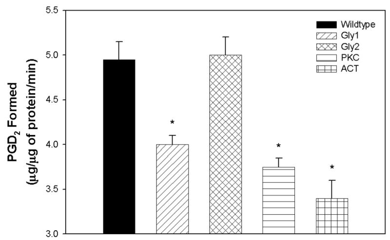
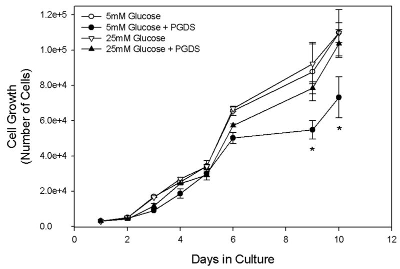
Lipocalin-type prostaglandin D(2) synthase (L-PGDS) is a highly glycosylated protein found in several body fluids. Elevated L-PGDS levels have been observed in the serum of patients with renal impairment, diabetes mellitus, and hypertension. Recently, we demonstrated the ability of L-PGDS to induce apoptosis in a variety of cell types including epithelial cells, neuronal cells, and vascular smooth muscle cells (VSMCs). The aim of this study was to investigate the effect several site-directed mutations had on L-PGDS-induced apoptosis in order to identify potential sites of regulation. Point mutations created in a glycosylation site (Asn51), a protein kinase C phosphorylation site (Ser106), and the enzymatic active site (Cys65) all inhibited L-PGDS-induced apoptosis as determined by both terminal deoxynucleotidyl transferase (TdT)-mediated dUTP nick end-labeling (TUNEL) and caspase3 activity. We also compared the L-PGDS isoforms present in GK rat serum to WKY control serum using two-dimensional gel electrophoresis and observed distinct differences which vanished after PNGase F glycolytic digestion. We conclude that post-translational modification of L-PGDS, by either glycosylation or phosphorylation, enhances its apoptotic activity and inhibits VSMC hyperproliferation and postulate that this process is altered in type 2 diabetes.
Figures
Similar articles
-
Prostaglandin D2 synthase inhibits the exaggerated growth phenotype of spontaneously hypertensive rat vascular smooth muscle cells.J Biol Chem. 2003 Jun 13;278(24):22175-81. doi: 10.1074/jbc.M302769200. Epub 2003 Apr 8. J Biol Chem. 2003. PMID: 12684506
-
Inhibition of cell cycle progression and migration of vascular smooth muscle cells by prostaglandin D2 synthase: resistance in diabetic Goto-Kakizaki rats.Am J Physiol Cell Physiol. 2004 Nov;287(5):C1273-81. doi: 10.1152/ajpcell.00230.2004. Epub 2004 Jul 7. Am J Physiol Cell Physiol. 2004. PMID: 15240344
-
Lipocalin-type prostaglandin D synthase protects against oxidative stress-induced neuronal cell death.Biochem J. 2012 Apr 1;443(1):75-84. doi: 10.1042/BJ20111889. Biochem J. 2012. PMID: 22248185
-
[Structure, localization and characterization of lipocalin-type prostaglandin D synthase].Zhonghua Nan Ke Xue. 2004 Feb;10(2):134-8. Zhonghua Nan Ke Xue. 2004. PMID: 15027190 Review. Chinese.
-
[Ligand recognition mechanism of lipocalin-type prostaglandin D synthase].Yakugaku Zasshi. 2011;131(11):1575-81. doi: 10.1248/yakushi.131.1575. Yakugaku Zasshi. 2011. PMID: 22041695 Review. Japanese.
Cited by
-
Lipocalin-type prostaglandin D2 synthase protein regulates glial cell migration and morphology through myristoylated alanine-rich C-kinase substrate: prostaglandin D2-independent effects.J Biol Chem. 2012 Mar 16;287(12):9414-28. doi: 10.1074/jbc.M111.330662. Epub 2012 Jan 24. J Biol Chem. 2012. PMID: 22275363 Free PMC article.
-
Distinct urinary glycoprotein signatures in prostate cancer patients.Oncotarget. 2018 Sep 4;9(69):33077-33097. doi: 10.18632/oncotarget.26005. eCollection 2018 Sep 4. Oncotarget. 2018. PMID: 30237853 Free PMC article.
-
Glycoprotein PTGDS promotes tumorigenesis of diffuse large B-cell lymphoma by MYH9-mediated regulation of Wnt-β-catenin-STAT3 signaling.Cell Death Differ. 2022 Mar;29(3):642-656. doi: 10.1038/s41418-021-00880-2. Epub 2021 Nov 6. Cell Death Differ. 2022. PMID: 34743203 Free PMC article.
-
Biochemical and Structural Characteristics, Gene Regulation, Physiological, Pathological and Clinical Features of Lipocalin-Type Prostaglandin D2 Synthase as a Multifunctional Lipocalin.Front Physiol. 2021 Oct 22;12:718002. doi: 10.3389/fphys.2021.718002. eCollection 2021. Front Physiol. 2021. PMID: 34744762 Free PMC article. Review.
-
Genome-wide transcription analysis of histidine-related cataract in Atlantic salmon (Salmo salar L).Mol Vis. 2009 Jul 9;15:1332-50. Mol Vis. 2009. PMID: 19597568 Free PMC article.
References
-
- Urade Y, Hayaishi O. Prostaglandin D synthase: structure and function. Vitam Horm. 2000;58:89–120. - PubMed
-
- Hiraoka A, Seiki K, Oda H, et al. Electrophoresis. 2001;22(16):3433–7. - PubMed
-
- Lescuyer P, Gandini A, Burkhard PR, Hochstrasser DF, Sanchez JC. Prostaglandin D2 synthase and its post-translational modifications in neurological disorders. Electrophoresis. 2005;26(23):4563–70. - PubMed
-
- Melegos DN, Freedman MS, Diamandis EP. Prostaglandin D synthase concentration in cerebrospinal fluid and serum of patients with neurological disorders. Prostaglandins. 1997;54(1):463–74. - PubMed
-
- Inoue T, Takayanagi K, Morooka S, et al. Thromb Haemost. 2001;85(1):165–70. - PubMed
Publication types
MeSH terms
Substances
Grants and funding
LinkOut - more resources
Full Text Sources
Molecular Biology Databases
