. 2007;35(3):999-1006.
doi: 10.1093/nar/gkl1124. Epub 2007 Jan 26.

Spectroscopic observation of RNA chaperone activities of Hfq in post-transcriptional regulation by a small non-coding RNA

Affiliations

Spectroscopic observation of RNA chaperone activities of Hfq in post-transcriptional regulation by a small non-coding RNA

Véronique Arluison et al. Nucleic Acids Res. 2007.

Abstract

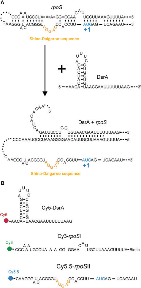
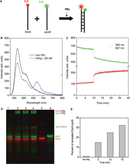
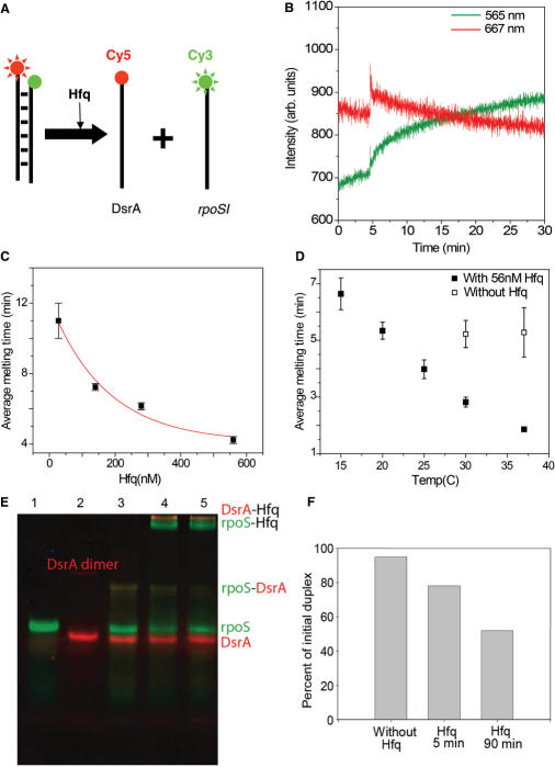
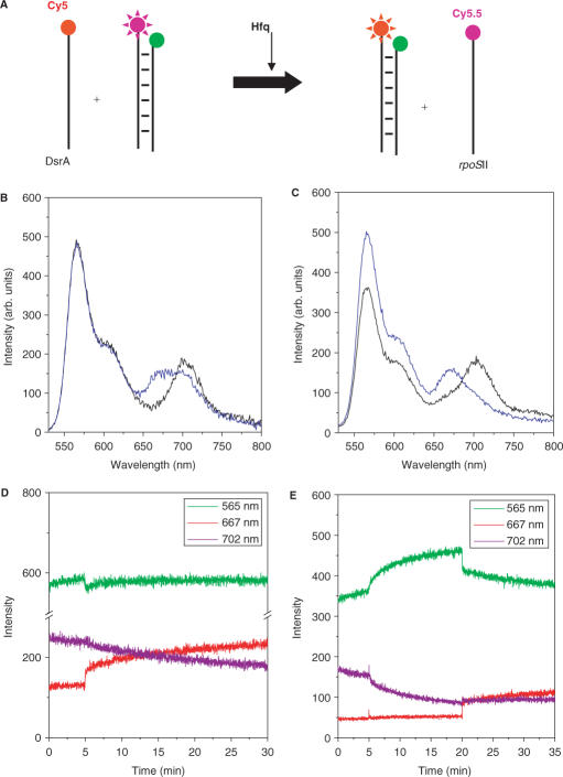
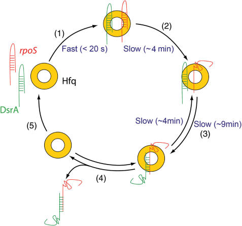
Hfq protein is vital for the function of many non-coding small (s)RNAs in bacteria but the mechanism by which Hfq facilitates the function of sRNA is still debated. We developed a fluorescence resonance energy transfer assay to probe how Hfq modulates the interaction between a sRNA, DsrA, and its regulatory target mRNA, rpoS. The relevant RNA fragments were labelled so that changes in intra- and intermolecular RNA structures can be monitored in real time. Our data show that Hfq promotes the strand exchange reaction in which the internal structure of rpoS is replaced by pairing with DsrA such that the Shine-Dalgarno sequence of the mRNA becomes exposed. Hfq appears to carry out strand exchange by inducing rapid association of DsrA and a premelted rpoS and by aiding in the slow disruption of the rpoS secondary structure. Unexpectedly, Hfq also disrupts a preformed complex between rpoS and DsrA. While it may not be a frequent event in vivo, this melting activity may have implications in the reversal of sRNA-based regulation. Overall, our data suggests that Hfq not only promotes strand exchange by binding rapidly to both DsrA and rpoS but also possesses RNA chaperoning properties that facilitates dynamic RNA-RNA interactions.


Figures

Figure 1.
Figure 1.
Translation regulation of rpoS by DsrA and FRET assay. (A) Proposed regulation mechanism of rpoS translation by DsrA. Base pairing between DsrA and rpoS 5′ UTR releases the rpoS segment containing the Shine–Dalgarno sequence. (B) RNA constructs for FRET measurements. DsrA, rpoSI and rpoSII are labelled with Cy5, Cy3 (and biotin) and Cy5.5, respectively.
Figure 2.
Figure 2.
Hfq helps annealing of DsrA and rpoS. (A) Scheme to examine the annealing of DsrA and rpoSI mediated by Hfq. (B) Emission spectra of 25 nM Cy3-rpoSI and 50 nM Cy5-DsrA in T50 buffer without Hfq or after incubating the sample for 10 min without Hfq (black line), and then with 28 nM of Hfq (blue line). (B) Emission intensity time trace of Cy3 at 565 nm (green trace) and Cy5 at 667 nm (red trace). Hfq (28 nM) added at 10 min results in an abrupt increase (<20s) in FRET followed by a slow increase (average annealing time = 9 min). (D) EMSA experiment confirms the annealing reaction. Green and red bands correspond to the fluorescence scans with Cy3 and Cy5 filters respectively. Lane 1: Cy3-rpoSI (25 nM) and Cy5-DsrA (50 nM) in the absence of Hfq, Lanes 2–4: Cy3-rpoSI and Cy5-DsrA in the presence of Hfq (28 nM) after incubation for 5, 10 and 15 min at 15°C, Lane 5: Cy5-DsrA and Lane 6: Cy3-rpoSI. (E) The amount of Cy3-RNA in the annealed form (indicated in the gel image) is quantified and shown in the plot.
Figure 3.
Figure 3.
Unwinding of DsrA+rpoS complex. (A) Schematic representation of rpoSI+DsrA melting by Hfq. (B) 25 nM rpoSI+DsrA complex was prepared in T50 buffer. The graph shows the intensity time traces of Cy3 at 565 nm (green line) and Cy5 at 667 nm (red lines). Hfq (28 nM) was added at 5 min. Increase in intensities of both dyes is followed by gradual decrease in FRET (average melting time = 11 min). (C) Average melting time as a function of Hfq concentration is plotted and fitted to an exponential decay function (red line). The fit decays to a value of 4.1 min for saturating concentrations of Hfq. (D) Average melting time as function of temperature in the presence of 56 nM Hfq (solid squares) and in the absence of Hfq (hollow squares) (E) An EMSA experiment that shows the melting reaction. Green and red bands correspond to the fluorescence scans with Cy3 and Cy5 filters respectively. Lane 1: Cy3-rpoSI (25 nM); Lane 2: Cy5-DsrA; Lane 3: Thermally annealed DsrA+rpoSI (25 nM); Lane 4–5: DsrA+rpoSI with 28 nM Hfq after 5 and 90 min. (F) The amount of Cy3-RNA in the annealed form is quantified and shown as a bar graph.
Figure 4.
Figure 4.
Hfq melts the stem structure of rpoS but does not promote annealing. (A) Scheme to show Hfq assisted melting of rpoS. (B) Emission spectra of 25 mM rpoSI+II or after incubating the sample for 5 min without Hfq (black line), and then for 25 min with 28 nM Hfq (blue line). (C) Intensity time trace of Cy3 (green lines) and Cy5.5 (red purple lines); Hfq (28 nM) was added at 5 min. Addition of Hfq results in gradual FRET decrease (average lifetime ∼15 min). (D) Average melting times were measured as a function of Hfq concentration (as plotted) and it decreases to ∼4 min in saturating concentrations of Hfq. (E) Hfq does not promote annealing of rpoSI and rpoSII. (F) Emission spectra of the mixture of 25 nM Cy3-rpoSI and 62.5 nM Cy5.5-rpoSII in T50 buffer without (black lines), after incubating the sample for 5 min without Hfq, and then with 28 nM of Hfq (blue lines).
Figure 5.
Figure 5.
Strand exchange reaction promoted by Hfq. (A) Schematic of the strand exchange reaction mediated by Hfq. (B) Emission spectra of the mixture of 25 nM of the rpoSI+II and 62.5 nM of Cy5-DsrA in T50 buffer without Hfq. Comparison of the emission spectra of the mixture 15 min after adding 28 nM Hfq (blue lines) and before adding Hfq (black lines) shows a shift in emission spectrum from Cy5.5 (707 nm) to Cy5 (667 nm). (C) Comparison of the emission spectra before (black line) and after addition of 282 nM Hfq (blue line). (D) Intensity time traces at Cy3, Cy5 and Cy5.5 emission wavelengths where 28 nM Hfq is added at t = 5 min to a solution containing rpoSI+II and Cy5-DsrA. (E) Intensity times traces at Cy3, Cy5 and Cy5.5 emission wavelengths where Hfq is added at t = 5 min and Cy5-DsrA is added at t = 20 min to a solution containing rpoSI+II.
Figure 6.
Figure 6.
A model for Hfq function in the translational regulation of rpoS by DsrA. (1) Hfq binds rapidly to both rpoS and DsrA. (2) After binding to rpoS, it slowly melts the stem region of rpoS. (3) Combined with the increased local concentration of both RNAs, this unwinding event could accelerate the annealing between rpoS and DsrA. Melting of the annealed DsrA … rpoS also takes place when bound to Hfq. (4) Hfq releases the DsrA … rpoS duplex and (5) it is free to get recycled.

References

    1. Franze de Fernandez MT, Hayward WS, August JT. Bacterial proteins required for replication of phage Qβ ribonucleic acid. J. Biol. Chem. 1972;247:824–821. - PubMed
    1. Vecerek B, Moll I, Blasi U. Translational autocontrol of the Escherichia coli hfq RNA chaperone gene. RNA. 2005;11:976–984. - PMC - PubMed
    1. Hajnsdorf E, Régnier P. Host factor HFq of Escherichia coli stimulates elongation of poly(A) tails by poly(A)polymerase I. Proc. Natl. Acad. Sc. U.S.A. 2000;97:1501–1505. - PMC - PubMed
    1. Rasmussen AA, Eriksen M, Gilany K, Udesen C, Franch T, Petersen C, Valentin-Hansen P. Regulation of ompA mRNA stability: the role of a small regulatory RNA in growth phase-dependent control. Mol. Microbiol. 2005;58:1421–1429. - PubMed
    1. Muffler A, Traulsen DD, Fischer D, Lange R, Hengge-Aronis R. The RNA-binding protein HF-1 plays a global regulatory role which is largely, but not exclusively, due to its role in expression of the σS subunit of RNA polymerase in Escherichia coli. J. Bacteriol. 1997;179:297–300. - PMC - PubMed

Publication types

MeSH terms