

Bacterial protein HU dictates the morphology of DNA condensates produced by crowding agents and polyamines
- PMID: 17259223
- PMCID: PMC1807954
- DOI: 10.1093/nar/gkl1093
Bacterial protein HU dictates the morphology of DNA condensates produced by crowding agents and polyamines
Abstract
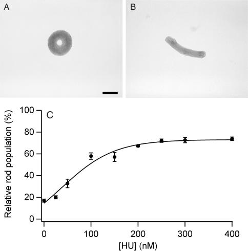
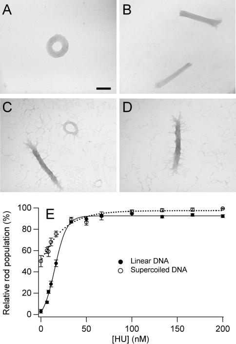
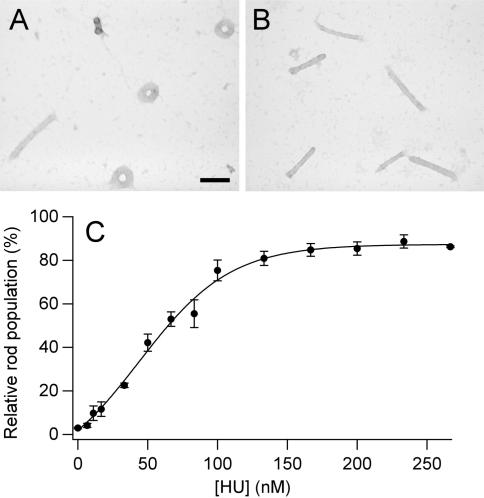
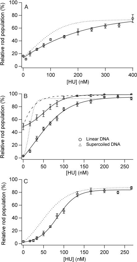
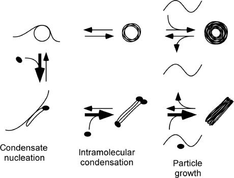
Controlling the size and shape of DNA condensates is important in vivo and for the improvement of nonviral gene delivery. Here, we demonstrate that the morphology of DNA condensates, formed under a variety of conditions, is shifted completely from toroids to rods if the bacterial protein HU is present during condensation. HU is a non-sequence-specific DNA binding protein that sharply bends DNA, but alone does not condense DNA into densely packed particles. Less than one HU dimer per 225 bp of DNA is sufficient to completely control condensate morphology when DNA is condensed by spermidine. We propose that rods are favored in the presence of HU because rods contain sharply bent DNA, whereas toroids contain only smoothly bent DNA. The results presented illustrate the utility of naturally derived proteins for controlling the shape of DNA condensates formed in vitro. HU is a highly conserved protein in bacteria that is implicated in the compaction and shaping of nucleoid structure. However, the exact role of HU in chromosome compaction is not well understood. Our demonstration that HU governs DNA condensation in vitro also suggests a mechanism by which HU could act as an architectural protein for bacterial chromosome compaction and organization in vivo.
Figures
Similar articles
-
Integration host factor (IHF) dictates the structure of polyamine-DNA condensates: implications for the role of IHF in the compaction of bacterial chromatin.Biochemistry. 2009 Feb 3;48(4):667-75. doi: 10.1021/bi8019965. Biochemistry. 2009. PMID: 19132923
-
Dual architectural roles of HU: formation of flexible hinges and rigid filaments.Proc Natl Acad Sci U S A. 2004 May 4;101(18):6969-74. doi: 10.1073/pnas.0308230101. Epub 2004 Apr 26. Proc Natl Acad Sci U S A. 2004. PMID: 15118104 Free PMC article.
-
Condensation of DNA by trivalent cations. 2. Effects of cation structure.Biopolymers. 1990;30(5-6):631-43. doi: 10.1002/bip.360300515. Biopolymers. 1990. PMID: 2265234
-
Architectural organization in E. coli nucleoid.Biochim Biophys Acta. 2012 Jul;1819(7):830-5. doi: 10.1016/j.bbagrm.2012.02.012. Epub 2012 Feb 22. Biochim Biophys Acta. 2012. PMID: 22387214 Free PMC article. Review.
-
DNA condensation in bacteria: Interplay between macromolecular crowding and nucleoid proteins.Biochimie. 2010 Dec;92(12):1715-21. doi: 10.1016/j.biochi.2010.06.024. Epub 2010 Jul 6. Biochimie. 2010. PMID: 20615449 Review.
Cited by
-
Effects of the nucleoid protein HU on the structure, flexibility, and ring-closure properties of DNA deduced from Monte Carlo simulations.J Mol Biol. 2008 Oct 3;382(2):353-70. doi: 10.1016/j.jmb.2008.05.088. Epub 2008 Jun 19. J Mol Biol. 2008. PMID: 18586040 Free PMC article.
-
Nucleoid-associated proteins shape chromatin structure and transcriptional regulation across the bacterial kingdom.Transcription. 2021 Aug;12(4):182-218. doi: 10.1080/21541264.2021.1973865. Epub 2021 Sep 9. Transcription. 2021. PMID: 34499567 Free PMC article. Review.
-
A histone-like protein induces plasmid DNA to form liquid crystals in vitro and gene compaction in vivo.Int J Mol Sci. 2013 Dec 6;14(12):23842-57. doi: 10.3390/ijms141223842. Int J Mol Sci. 2013. PMID: 24322443 Free PMC article.
-
Extracellular DNA-protein interactions.Curr Opin Struct Biol. 2024 Dec;89:102943. doi: 10.1016/j.sbi.2024.102943. Epub 2024 Oct 16. Curr Opin Struct Biol. 2024. PMID: 39418796 Review.
-
Understanding apparent DNA flexibility enhancement by HU and HMGB architectural proteins.J Mol Biol. 2011 Jun 3;409(2):278-89. doi: 10.1016/j.jmb.2011.03.050. Epub 2011 Apr 1. J Mol Biol. 2011. PMID: 21459097 Free PMC article.
References
-
- Gosule LC, Schellman JA. Compact form of DNA induced by spermidine. Nature. 1976;259:333–335. - PubMed
-
- Bloomfield V. DNA Condensation. Curr. Opin. Struct. Biol. 1996;6:334–341. - PubMed
-
- Bloomfield V. DNA condensation by multivalent cations. Biopolymers. 1997;44:269–282. - PubMed
-
- Hud NV, Vilfan ID. Toroidal DNA condensates: unraveling the fine structure and the role of nucleation in determining size in vitro. Annu. Rev. Biophys. Biomol. Struct. 2005;34:295–318. - PubMed
Publication types
MeSH terms
Substances
Grants and funding
LinkOut - more resources
Full Text Sources
Other Literature Sources
Molecular Biology Databases
