. 2007 Jan;19(1):351-68.
doi: 10.1105/tpc.106.048033. Epub 2007 Jan 26.

The acyltransferase GPAT5 is required for the synthesis of suberin in seed coat and root of Arabidopsis

Affiliations

The acyltransferase GPAT5 is required for the synthesis of suberin in seed coat and root of Arabidopsis

Fred Beisson et al. Plant Cell. 2007 Jan.

Abstract

Suberin and cutin are fatty acid- and glycerol-based plant polymers that act as pathogen barriers and function in the control of water and solute transport. However, despite important physiological roles, their biosynthetic pathways, including the acyl transfer reactions, remain hypothetical. We report the characterization of two suberin mutants (gpat5-1 and gpat5-2) of Arabidopsis thaliana GPAT5, encoding a protein with acyl-CoA:glycerol-3-phosphate acyltransferase activity. RT-PCR and beta-glucuronidase-promoter fusion analyses demonstrated GPAT5 expression in seed coat, root, hypocotyl, and anther. The gpat5 plants showed a 50% decrease in aliphatic suberin in young roots and produced seed coats with a severalfold reduction in very long chain dicarboxylic acid and omega-hydroxy fatty acids typical of suberin but no change in the composition or content of membrane or storage glycerolipids or surface waxes. Consistent with their altered suberin, seed coats of gpat5 mutants had a steep increase in permeability to tetrazolium salts compared with wild-type seed coats. Furthermore, the germination rate of gpat5 seeds under high salt was reduced, and gpat5 seedlings had lower tolerance to salt stress. These results provide evidence for a critical role of GPAT5 in polyester biogenesis in seed coats and roots and for the importance of lipid polymer structures in the normal function of these organs.


Figures

Figure 1.
Figure 1.
Structure of the GPAT5 Gene Carrying a T-DNA Insertion, and Analysis of GPAT5 Expression by RT-PCR. (A) Genomic organization of the gpat5-1 and gpat5-2 loci. Boxes represent exons. The T-DNA insertion point is indicated as a triangle, with L and R indicating left and right borders, respectively. (B) RT-PCR analysis of the GPAT5 transcript in wild-type and mutant (gpat5-1 and gpat5-2) flowers. Approximately 0.1 μg of total RNA was used in each PCR, and eIF4A-1 (At3g13920) was used as a control. (C) RT-PCR analysis of GPAT5 expression in roots, rosette leaves, stems, open flowers, and developing seeds. Approximately 0.1 μg of total RNA was used in each PCR, and eIF4A-1 (At3g13920) was used as a control.
Figure 2.
Figure 2.
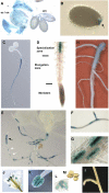
Analysis of GPAT5 Expression in Arabidopsis Wild-Type Plants by ProGPAT5:GUS. (A) and (B) Crushed GUS-stained seeds show that staining is limited to the seed coat/endosperm. The beginning (A) and end (B) stages of seed desiccation are shown. In (B), staining is limited to the funiculus attachment region only (arrow). em, embryo; en, endosperm; sc, seed coat. (C) GUS staining in a 4-d-old seedling grown on agar. (D) GUS staining is detected in the specialization zone but not in the meristem and elongation zone of the root in a 7-d-old seedling. (E) GUS staining of a 3-week-old seedling. For arrows, see text. (F) GUS staining in a seminal root and a first-order lateral root of a 3-week-old seedling. (G) Patchy GUS staining is often observed in roots of 1- to 4-week-old seedlings. (H) GUS staining is faint in roots of 6-week-old plants on soil and is restricted to the older parts. (I) GUS staining is not observed in stems and cauline leaves. (J) GUS staining in flowers is restricted to the stamen. (K) GUS staining is observed in anthers but not in filaments of the stamen. (L) GUS staining is observed in developing pollen. (M) GUS staining is not observed in mature pollen.
Figure 3.
Figure 3.
Fatty Acids from the Seed Coat/Endosperm Fraction of the Wild Type and gpat5 Mutants. Mature seeds were manually dissected, and total fatty acids of the membrane and storage lipids of the seed coat/endosperm fraction were analyzed as fatty acid methyl esters by gas chromatography. Values are means of six replicates. Error bars denote 95% confidence intervals.
Figure 4.
Figure 4.
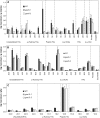
Lipid Polyester Monomers from Seeds, Roots, and Flowers of Wild-Type and gpat5 Plants. (A) Polyester monomers from mature seeds. (B) Polyester monomers from roots of 1-week-old seedlings grown on agar. (C) Polyester monomers from opened flowers. The insoluble dry residue obtained after grinding and delipidation of tissues with organic solvents was depolymerized with sodium methoxide, and aliphatic and aromatic monomers released were analyzed by gas chromatography–mass spectrometry. Values are means of six data points (two independent experiments using different biological samples involving triplicate assays for the depolymerization reaction). Error bars denote 95% confidence intervals. DCAs, fatty dicarboxylic acids; FAs, fatty acids; fw, fresh weight; PAs, primary alcohols. The polyol fatty acids are 10,16-hydroxy 16:0 and 9,10,18-hydroxy 18:1.
Figure 5.
Figure 5.
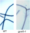
Staining of 3-Week-Old Roots of gpat5-1 and Wild-Type Plants with the Suberin Dye Sudan Black B.
Figure 6.
Figure 6.
Phenotype of gpat5 Seeds, and Permeability to Dyes. Similar results were obtained with gpat5-2. (A) Scanning electron microscopy of the surface of wild-type versus gpat5-1 seeds. Bars = 40 μm. (B) Ruthenium red staining of seed mucilage in wild-type versus gpat5-1 seeds. (C) Tetrazolium salt staining (24 h) of wild-type versus gpat5-1 seeds. (D) Seeds from (C) dissected after staining. (E) Tetrazolium salt staining (4 h) of wild-type versus gpat5-1 seeds. (F) Autofluorescence of wild-type versus gpat5-1 seeds (365-nm excitation). (G) Sudan red 7B staining of wild-type versus gpat5-1 seeds. (H) Tetrazolium salt staining (24 h) of seeds resulting from the fertilization of wild-type plants by gpat5-1 pollen (left) and of gpat5-1 plants by wild-type pollen (right).
Figure 7.
Figure 7.
Brown Pigmentation of Wild-Type and gpat5 Seeds. (A) Batch color of wild-type versus gpat5-1 and gpat5-2 seeds. (B) Amount of soluble and insoluble PAs in wild-type versus gpat5-1 and gpat5-2 seeds. Values are means of six data points (two independent experiments using different seed batches involving triplicate assays for the depolymerization reactions). Error bars denote 95% confidence intervals.
Figure 8.
Figure 8.
Germination of gpat5 Seeds under Various Conditions. (A) Rate of germination after harvest and increasing periods of dry storage. (B) Rate of germination on MS medium supplemented with increasing NaCl concentrations. (C) Rate of germination on MS medium supplemented with increasing KCl concentrations. (D) Rate of germination on MS medium supplemented with increasing K2SO4 concentrations. Seeds were germinated after cold treatment (except in [A]). Values are means of 9 data points (A) or 12 data points ([B] to [D]; i.e., from three or four independent experiments, respectively, which use different seed batches and involve three replicate lots of ∼100 seeds for each seed batch). Error bars denote 95% confidence intervals. Similar results were obtained with gpat5-2.
Figure 9.
Figure 9.
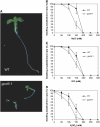
Rate of Seedling Establishment under Increasing Salt Concentrations, and Phenotypes of Seedlings Germinated on 150 mM NaCl. (A) Seedlings after 12 d at 150 mM NaCl. All seedlings were photographed at the same magnification. (B) Rate of seedling establishment under increasing NaCl concentrations. (C) Rate of seedling establishment under increasing KCl concentrations. (D) Rate of seedling establishment under increasing K2SO4 concentrations. After cold treatment, seeds were germinated for 12 d and seedling establishment was scored. Values are means of 12 data points (four independent experiments using different seed batches involving three replicate lots of ∼100 seeds for each seed batch). Error bars denote 95% confidence intervals. Similar results were obtained with gpat5-2.
Figure 10.
Figure 10.
Rate of Seedling Bleaching at 5 d after Transfer on 200 mM NaCl. Seeds were germinated on MS medium for 3 d and transferred to MS medium supplemented with 200 mM NaCl. Values are means of nine data points (three independent experiments using different seed batches involving three replicate lots of ∼100 seeds for each seed batch). Error bars denote 95% confidence intervals.
Figure 11.
Figure 11.
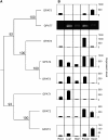
Gene Tree and Gene Expression Profiles of the Eight Putative GPATs of Arabidopsis. (A) The cladogram shows the branching order of Arabidopsis GPATs according to a phylogenetic tree of protein sequences of plant acyltransferases (Kim and Huang, 2004). The original tree was built using the neighbor-joining method with 1000 bootstrap replicates. Bootstrap values are percentages. (B) Microarray expression data derived from AtGenExpress (Schmid et al., 2005). Expression levels in each tissue (root, leaf, stem, flower, and seed) at different developmental stages were averaged (bars represent means ± se). The expression profile for GPAT7 was determined in this study via RT-PCR analysis because its expression profile is not available at AtGenExpress.

References

    1. Alonso, J.M., et al. (2003). Genome-wide insertional mutagenesis of Arabidopsis thaliana. Science 301 653–657. - PubMed
    1. Baum, S.F., Dubrovsky, J.G., and Rost, L.T. (2002). Apical organization and maturation of the cortex and vascular cylinder in Arabidopsis thaliana (Brassicaceae) roots. Am. J. Bot. 89 908–920. - PubMed
    1. Bechtold, N., Ellis, J., and Pelletier, G. (1993). In planta Agrobacterium-mediated gene transfer by infiltration of adult Arabidopsis thaliana plants. C. R. Acad. Sci. 316 1194–1199. - PubMed
    1. Beeckman, T., De Rycke, R., Viane, R., and Inze, D. (2000). Histological study of seed coat development in Arabidopsis thaliana. J. Plant Res. 113 139–148.
    1. Beisson, F., et al. (2003). Arabidopsis genes involved in acyl lipid metabolism. A 2003 census of the candidates, a study of the distribution of expressed sequence tags in organs, and a web-based database. Plant Physiol. 132 681–697. - PMC - PubMed

Publication types

MeSH terms