. 2007 Feb 20;104(8):2571-8.
doi: 10.1073/pnas.0609498104. Epub 2007 Jan 26.

Hepatitis B virus X protein enhances androgen receptor-responsive gene expression depending on androgen level

Affiliations

Hepatitis B virus X protein enhances androgen receptor-responsive gene expression depending on androgen level

Chi-Ming Chiu et al. Proc Natl Acad Sci U S A. .

Abstract

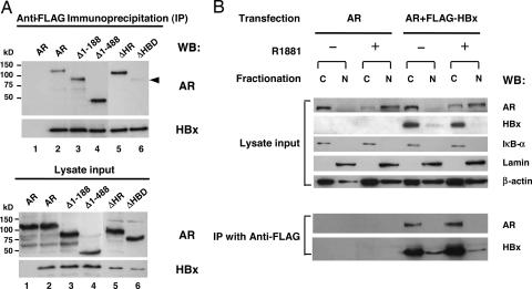
Persistent hepatitis B virus (HBV) infection is a major risk of hepatocellular carcinoma (HCC). One intriguing feature of HBV-related HCC is the male predominance, with a male to female ratio of 5-7:1. This dominance has been attributed to the elevated androgen level and the enhanced androgen receptor (AR)-mediated activity in the host. How HBV infection and AR signaling modulate HCC is unknown. We investigated whether the HBV nonstructural protein, X protein (HBx) could cooperate with the AR signaling pathway to enhance carcinogenesis. We found that HBx increased the anchorage-independent colony-formation potency of AR in a nontransformed mouse hepatocyte cell line. We also found that HBx functioned as a positive transcriptional coregulator to increase AR-mediated transcriptional activity. This transcription enhancement was increased in the presence of androgen in a concentration-responsive manner, thus explaining a more prominent effect in males. HBx did not physically associate with ligand-bound AR in the nucleus, and it likely augmented AR activity by increasing the phosphorylation of AR through HBx-mediated activation of the c-Src kinase signaling pathway. Our study documents HBx as a previously undescribed class of noncellular positive coregulators for AR. The results reveal a mechanism for the vulnerability of males to microbial infections and the subsequent development of cancer.


Conflict of interest statement

The authors declare no conflict of interest.

Figures

Fig. 1.
Fig. 1.
HBx augments androgen-dependent AR-responsive gene expression. (A) HepG2 cells were transfected with pMMTV-luc reporter, pRL-CMV, pSG5-AR, and pCMV-HBx with different combinations. Cells were then treated with DHT with concentrations as indicated, and the cell lysates were prepared for assessment of luciferase activity. The results shown are the average of three experiments (mean ± SD). (B) The experiment was similar to the above, except that Huh-7 cells were used, pCMV-HBx was replaced with either pHBV-48 or pHBV-48-X0, and DHT was replaced with 10 nM R1881. The results shown are the average of three experiments (mean ± SD). (C) Western blot analysis was used to verify protein expression by probing with antibodies as indicated. HBc, hepatitis B core antigen.
Fig. 2.
Fig. 2.
Functional characterization of HBx and AR regions responsible for HBx-induced AR activation. (A) Schematic representation of the four site-directed mutants of HBx used for transfection experiments. (B) Similar to Fig. 1A, except that pSG5-AR was cotransfected with wild-type or site-directed mutants of pSV-HBx, and 10 nM R1881 was added. The results shown are the average of three experiments (mean ± SD). (C) Schematic illustration of wild type and the four deletion constructs of AR used for the transfection experiments with the functional domains and the deletion regions indicated. (D) The expression of AR constructs in transfected Huh-7 cells was analyzed by immunoblotting. Protein bands corresponding to AR constructs are indicated by the arrowheads. (E) Huh-7 cells were transfected with plasmids as indicated, either treated or untreated with 10 nM R1881, and luciferase activities were determined. Results are the average of three experiments (mean ± SD).
Fig. 3.
Fig. 3.
Characterization of the interaction between AR and HBx and their subcellular distribution pattern in Huh-7 cells. (A Upper) Cell lysates from Huh-7 cells transfected with FLAG-HBx and the AR constructs were processed for coimmunoprecipitation analysis, and the immunoprecipitates were analyzed by Western blot analysis probed with AR and HBx antibodies. The position of ARΔHBD protein is denoted by the arrowhead. (Lower) Expression of HBx and AR proteins. (B) Huh-7 cells were transfected with pSG5-AR and FLAG-HBx, treated with 10 nM R1881 or ethanol, and fractionated into cytosolic and nuclear fractions. (Upper) Fifty micrograms of the extracts was loaded as lysate input for Western blot analysis by using antibodies for AR and HBx. IκBα antibody was used as the cytosolic protein control, and lamin B antibody was used as the nuclear protein control. (Lower) Two hundred fifty micrograms of lysates was used for immunoprecipitation and Western blot analysis by using AR and HBx antibodies.
Fig. 4.
Fig. 4.
Involvement of c-Src activity in HBx induced AR activation and AR phosphorylation. (A) HepG2 cells cotransfected with pSG5-AR, pCMV-HBx, pMMTV-luc, and pRL-CMV were untreated, treated with 10 nM R1881, or treated with a combination of the inhibitors PP2 (20 μM), U0126 (10 μM), LY294002 (100 nM), SP600125 (20 nM), and cyclosporine A (10 μg/ml), as indicated. (B) HepG2 cells were transfected with pSG5-AR, pCMV-HBx, pMMTV-Luc, pRL-CMV, and the dominant-negative c-Src mutant K295M or the CSK kinase expression plasmids, and treated with 10 nM R1881 as indicated. Luciferase activities are the average of three experiments (mean ± SD). (C) Huh-7 cells transfected with pSG5-AR and pCMV-HBx were treated with R1881 (10 nM) in combination with PP2 (20 or 50 μM). The cell lysates were harvested for Western blot analysis by using the indicated antibodies. The p-Y418Src antibody was used as a control to verify the suppression of c-Src kinase activity by PP2 treatment.
Fig. 5.
Fig. 5.
A proposed model for HBx induced AR activation and carcinogenesis. HBx-mediated enhancement of AR activity is androgen-dependent and could be mediated through an indirect mechanism involving calcium and c-Src signaling pathways, which lead to subsequent augmentation of AR phosphorylation and increased transcriptional activities (the genomic effect). Alternatively, nongenomic effects mediated by c-Src signaling in the cytoplasm might affect cell proliferation and survival (indicated as gray characters), although the present study did not explore this possibility. The details of this proposed model are discussed in the text.

Similar articles

Cited by

References

    1. Parkin DM, Bray F, Ferlay J, Pisani P. Int J Cancer. 2001;94:153–156. - PubMed
    1. Chen CJ, Yu MW, Liaw YF. J Gastroenterol Hepatol. 1997;12:S294–308. - PubMed
    1. Yu MW, Chen CJ. Crit Rev Oncol Hematol. 1994;17:71–91. - PubMed
    1. De Maria N, Manno M, Villa E. Mol Cell Endocrinol. 2002;193:59–63. - PubMed
    1. Kemp CJ, Leary CN, Drinkwater NR. Proc Natl Acad Sci USA. 1989;86:7505–7509. - PMC - PubMed

Publication types

MeSH terms

LinkOut - more resources