

Review
doi: 10.1128/EC.00381-06.
Epub 2007 Jan 26.
Hyphal growth: a tale of motors, lipids, and the Spitzenkörper
Affiliations
- PMID: 17259546
- PMCID: PMC1828937
- DOI: 10.1128/EC.00381-06
Item in Clipboard
Review
Hyphal growth: a tale of motors, lipids, and the Spitzenkörper
Eukaryot Cell.
2007 Mar.
No abstract available
Figures

Tip growth and the Spitzenkörper in Neurospora crassa strain 74A. Rapid hyphal elongation through 1% low-melting agarose is accompanied by a dynamic accumulation of vesicles in the apex (dark in phase-contrast micrographs, blue in pseudo-colored image). Elapsed time is given in minutes:seconds. Bar, 10 μm. The images are taken from Movie S1 in the supplemental material.

GFP-labeled myosin-5 in a hypha of a myo5 null mutant of Ustilago maydis. The functional fusion protein concentrates in the hyphal tip, where it is dynamically rearranged. Note that faint GFP-Myo5 signals show directed motility in the subapical regions, which is best seen in the movie available at http://www.mpi-marburg.mpg.de/downloads/ . Shown is strain AB33ΔMyo5_GFP3Myo5, in which the the b-transcription factor that triggers filamentous growth is under the control of inducible promoters (14). In addition, the endogenous myo5 gene is deleted and a triple GFP fused to Myo5 is ectopically integrated. Elapsed time is given in seconds. Bars, 5 and 2 μm.
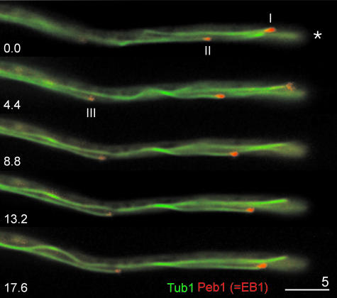
Orientation of microtubules in a hypha of Ustilago maydis. A functional fusion protein of the EB1-homologue Peb1 and RFP binds to the plus end of elongating microtubules (indicated by roman numbers). About 90% of all plus ends are directed to the hyphal tip (indicated by an asterisk). Elapsed time is given in seconds. Bar, 5 μm. Shown is strain AB33Peb1RFP_GT, in which MTs are labeled by ectopically expressed GFP-α-tubulin, and which contains the endogenous plus end-binding EB1-homologue Peb1 (121) fused to RFP. The images are taken from a movie available at http://www.mpi-marburg .mpg.de/downloads/.

Motility of early endosomes along microtubules in Ustilago maydis. Organelles are marked by Yup1, a tSNARE that locates to early endosomes (134). In hyphae, endosomes (Yup1-GFP; green) move along RFP-α-tubulin-containing microtubules (Tub1; red) to the tip, where they remain for a short period and return to subapical regions (arrow) (67). Elapsed time is given in seconds. Bar, 5 μm. Shown is strain AB33Yup1GFP_RT, in which MTs are labeled by ectopicall expressed GFP-α-tubulin and which contains an ectopically expressed endosomal tSNARE Yup1 fused to GFP (134). The images are taken from Movie S2 in the supplemental material.

Motility of a wall-less Neurospora crassa mutant cell in phosphate-buffered saline. Cells taken from solid medium show amoeboid movement in osmotically stabilizing buffer. The formation of cellular extensions in the absence of a cell wall indicates that internal cytoplasmic forces are generated that might involve the cytoskeleton. Elapsed time is given in minutes:seconds. Bar, 5 μm. Shown is strain fz;sg;os-1 (“slime”; FGSC 1118), which is defective in (1-3)-β-d -glucan synthase. The images were taken from the movie available at http://www.mpi-marburg.mpg.de/downloads/ .

Motility of a wall-less Neurospora crassa mutant cell attached to a protamine sulfate-coated coverslip. The cell is able to push out cytoplasmic extensions that remain attached and thereby “crawls” over the surface (arrow and arrowhead). Elapsed time is given in minutes:seconds. Bar, 5 μm. Shown is strain fz;sg;os-1 (“slime”; FGSC 1118), which defective in (1-3)-β-d -glucan synthase. The images were taken from the Movie S3 in the supplemental material.
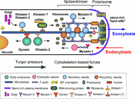
A model of hyphal tip growth. Sterol-rich membrane domains are concentrated at the hyphal apex, where they might facilitate endocytosis. Myosin-1 participates in initial steps of endocytosis by polymerizing actin, which “rockets” vesicles away from the membrane. In addition, lipid rafts could provide a scaffold for apical proteins, such as phosphatidylinositol-4-phosphate 5-kinase (PIP5K) (62), that generate a functional microdomain, which might recruit components of the polarisome (Bud6 and Spa2) and Cdc42 (20, 61). Spa2 and Bud6 bind formins (26, 35), which themselves participate in nucleation and polarization of the F-actin cytoskeleton (64, 95). Consequently, tip-ward delivery of exocytic vesicles becomes possible. MTs might be anchored at the tip via kinesin-motors (63), which supports long-distance kinesin-1- and kinesin-3-based transport (108) that delivers vesicles, but also might take mRNA to the tip (11), where translation at apical ribosomes (40, 56) and further processing in the endoplasmic reticulum and the apical Golgi (103) happen. Note that the endoplasmic reticulum also concentrates in the hyphal tip (135), but was left out for simplicity. Short-distance transport might be mediate by myosin-5 motors (108, 133). Vesicles are stored in the Spitzenkörper, which determines hyphal growth by controlled exocytosis. Cytoplasmic forces that could be generated by turgor pressure and cytoskeleton-based dynamics, both pushing the flexible apex forward. Note that the presented model is based on data from several model systems, including Fusarium acuminatum, S. cerevisiae, Ashbya gossypii, A. nidulans, C. albicans, and U. maydis.
References
-
- Agrios, G. N. 1997. Plant pathology. Academic Press, London, United Kingdom.
-
- Akashi, T., T. Kanbe, and K. Tanaka. 1994. The role of the cytoskeleton in the polarized growth of the germ tube in Candida albicans. Microbiology 140:271-280. - PubMed
-
- Archer, D. B., and D. A. Wood. 1995. Fungal exoenzymes, p. 135-162. In N. A. R. Gow and G. M. Gadd (ed.), The growing fungus. Chapman and Hall, London, United Kingdom.
Publication types
MeSH terms
Substances
LinkOut - more resources
Full Text Sources
Medical
