Developmental changes and injury induced disruption of the radial organization of the cortex in the immature rat brain revealed by in vivo diffusion tensor MRI
- PMID: 17259644
- PMCID: PMC4780675
- DOI: 10.1093/cercor/bhl168
Developmental changes and injury induced disruption of the radial organization of the cortex in the immature rat brain revealed by in vivo diffusion tensor MRI
Abstract
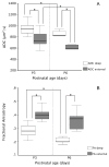
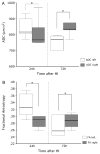
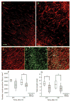
During brain development, morphological changes modify the cortex from its immature radial organization to its mature laminar appearance. Applying in vivo diffusion tensor imaging (DTI), the microstructural organization of the cortex in the immature rat was analyzed and correlated to neurohistopathology. Significant differences in apparent diffusion coefficient (ADC) and fractional anisotropy (FA) were detected between the external (I-III) and deep (IV-VI) cortical layers in postnatal day 3 (P3) and P6 pups. With cortical maturation, ADC was reduced in both cortical regions, whereas a decrease in FA was only seen in the deep layers. A distinct radial organization of the external cortical layers with the eigenvectors perpendicular to the pial surface was observed at both ages. Histology revealed maturational differences in the cortical architecture with increased neurodendritic density and reduction in the radial glia scaffolding. Early DTI after hypoxia-ischemia at P3 shows reduced ADC and FA in the ipsilateral cortex that persisted at P6. Cortical DTI eigenvector maps reveal microstructural disruption of the radial organization corresponding to regions of neuronal death, radial glial disruption, and astrocytosis. Thus, the combined use of in vivo DTI and histopathology can assist in delineating normal developmental changes and postinjury modifications in the immature rodent brain.
Figures


Similar articles
-
In vivo high-resolution diffusion tensor imaging of the developing neonatal rat cortex and its relationship to glial and dendritic maturation.Brain Struct Funct. 2019 Jun;224(5):1815-1829. doi: 10.1007/s00429-019-01878-w. Epub 2019 Apr 22. Brain Struct Funct. 2019. PMID: 31011813 Free PMC article.
-
Cellular correlates of longitudinal diffusion tensor imaging of axonal degeneration following hypoxic-ischemic cerebral infarction in neonatal rats.Neuroimage Clin. 2014 Aug 7;6:32-42. doi: 10.1016/j.nicl.2014.08.003. eCollection 2014. Neuroimage Clin. 2014. PMID: 25379414 Free PMC article.
-
Imaging and serum biomarkers reflecting the functional efficacy of extended erythropoietin treatment in rats following infantile traumatic brain injury.J Neurosurg Pediatr. 2016 Jun;17(6):739-55. doi: 10.3171/2015.10.PEDS15554. Epub 2016 Feb 19. J Neurosurg Pediatr. 2016. PMID: 26894518 Free PMC article.
-
The role of diffusion tensor imaging in the evaluation of ischemic brain injury - a review.NMR Biomed. 2002 Nov-Dec;15(7-8):561-9. doi: 10.1002/nbm.786. NMR Biomed. 2002. PMID: 12489102 Review.
-
The role of diffusion tensor imaging and fractional anisotropy in the evaluation of patients with idiopathic normal pressure hydrocephalus: a literature review.Neurosurg Focus. 2016 Sep;41(3):E12. doi: 10.3171/2016.6.FOCUS16192. Neurosurg Focus. 2016. PMID: 27581308 Review.
Cited by
-
Connectome and Maturation Profiles of the Developing Mouse Brain Using Diffusion Tensor Imaging.Cereb Cortex. 2015 Sep;25(9):2696-706. doi: 10.1093/cercor/bhu068. Epub 2014 Apr 6. Cereb Cortex. 2015. PMID: 24711485 Free PMC article.
-
Regional microstructural organization of the cerebral cortex is affected by preterm birth.Neuroimage Clin. 2018 Mar 16;18:871-880. doi: 10.1016/j.nicl.2018.03.020. eCollection 2018. Neuroimage Clin. 2018. PMID: 29876271 Free PMC article.
-
Unbiased Quantification of Subplate Neuron Loss following Neonatal Hypoxia-Ischemia in a Rat Model.Dev Neurosci. 2017;39(1-4):171-181. doi: 10.1159/000460815. Epub 2017 Apr 22. Dev Neurosci. 2017. PMID: 28434006 Free PMC article.
-
Diffusion kurtosis imaging: An efficient tool for evaluating age-related changes in rat brains.Brain Behav. 2021 Nov;11(11):e02136. doi: 10.1002/brb3.2136. Epub 2021 Sep 24. Brain Behav. 2021. PMID: 34559478 Free PMC article.
-
Determination of axonal and dendritic orientation distributions within the developing cerebral cortex by diffusion tensor imaging.IEEE Trans Med Imaging. 2012 Jan;31(1):16-32. doi: 10.1109/TMI.2011.2162099. Epub 2011 Jul 14. IEEE Trans Med Imaging. 2012. PMID: 21768045 Free PMC article.
References
-
- Back SA, Kinney HC, Volpe JJ. Immunocytochemical characterization of oligodendrocyte development in
-
- Baratti C, Barnett AS, Pierpaoli C. Comparative MR imaging study of brain maturation in kittens with T1 - PubMed
-
- Basser PJ, Pierpaoli C. Microstructural and physiological features of tissues elucidated by quantitative-diffusion-tensor MRI. J Magn Reson B. 1996;111:209–219. - PubMed
-
- Beaulieu C, Allen PS. Determinants of anisotropic water diffusion in nerves. Magn Reson Med. 1994;31:394–400. - PubMed
-
- Brazel CY, Romanko MJ, Rothstein RP, Levison SW. Roles of the mammalian subventricular zone in brain development. Prog Neurobiol. 2003;69:49–69. - PubMed
Publication types
MeSH terms
Grants and funding
LinkOut - more resources
Full Text Sources
Medical
Research Materials

