. 2006:2006:69612.
doi: 10.1155/PPAR/2006/69612.

Examination of Ligand-Dependent Coactivator Recruitment by Peroxisome Proliferator-Activated Receptor-alpha (PPARalpha)

Affiliations

Examination of Ligand-Dependent Coactivator Recruitment by Peroxisome Proliferator-Activated Receptor-alpha (PPARalpha)

Eric S Tien et al. PPAR Res. 2006.

Abstract

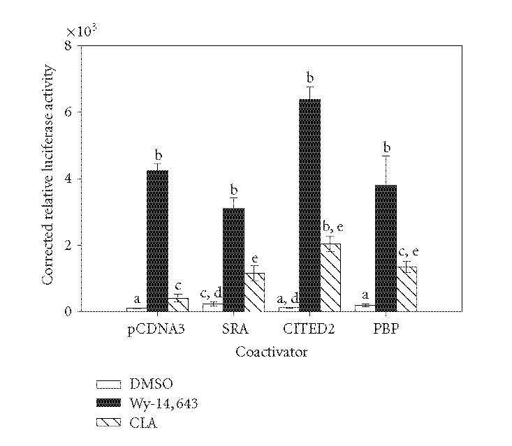
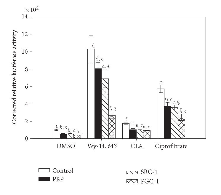
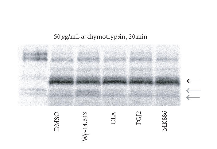
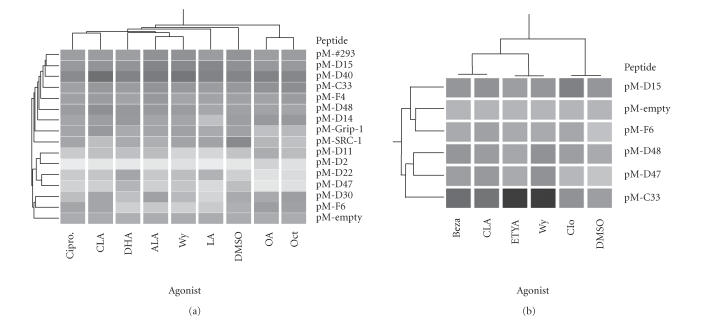
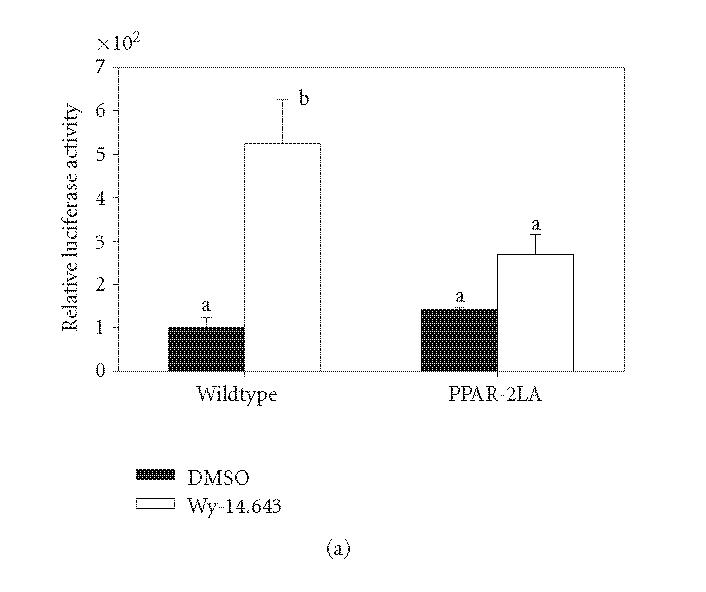
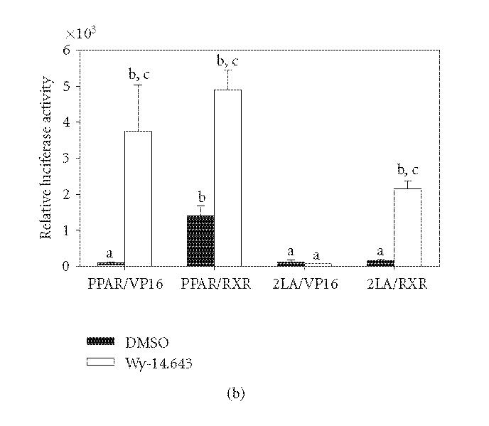
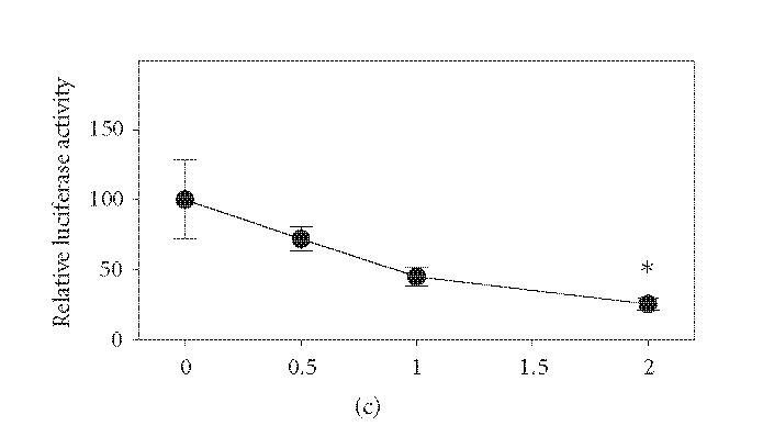
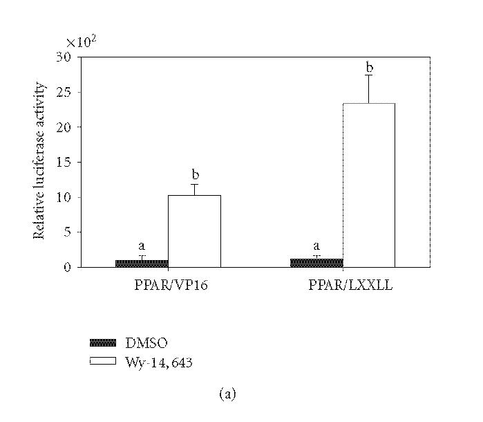
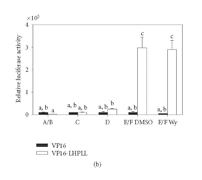
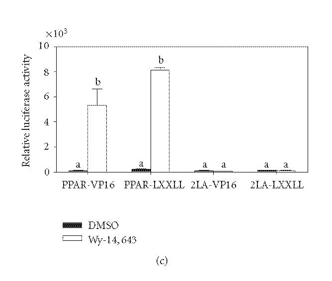
The ligand-dependent recruitment of coactivators to peroxisome proliferator-activated receptor-alpha (PPARalpha) was examined. PPAR-binding protein (PBP), PPARgamma coactivator-1alpha (PGC-1alpha), steroid receptor coactivator-1 (SRC-1), and CBP/p300-interacting transactivator with ED-rich tail 2 (CITED2) affected PPARalpha activity in the presence of Wy-14,643. The effects on PPARalpha activity in light of increased or decreased expression of these coactivators were qualitatively different depending on the ligand examined. Diminished expression of PGC-1alpha, SRC-1, or PBP by RNAi plasmids affected natural or synthetic agonist activity whereas only Wy-14,643 was affected by decreased PGC-1alpha. The interaction of PPARalpha with an LXXLL-containing peptide library showed ligand-specific patterns, indicative of differences in conformational change. The association of coactivators to PPARalpha occurs predominantly via the carboxyl-terminus and mutating (456)LHPLL to (456)LHPAA resulted in a dominant-negative construct. This research confirms that coactivator recruitment to PPARalpha is ligand-dependent and that selective receptor modulators (SRMs) of this important protein are likely.


Figures

Figure 1
Figure 1
Effects of various coactivators on the activation of PPARα by Wy-14,643 and CLA. COS-1 cells were transiently transfected with pM/PPARα, GAL4 reporter, and coactivator expression plasmids or empty vector control (total DNA per well was held constant). Cells were treated for 6 hours with 50 μM Wy-14,643, 100 μM CLA, or DMSO. Luciferase activity was measured and corrected for transfection efficiency and extraction yield (n = 3). Relative values are corrected to untreated pcDNA3 bar (100%). Bars with different letters above are significantly different from each other (Tukey's multicomparison test, P < .05), representative of 2 independent experiments.
Figure 2
Figure 2
Inhibition of coactivator expression by RNAi decreases PPARα activity in a ligand-specific manner. 3T3L1 cells were transiently transfected with pM/PPARα, GAL4 responsive reporter, pRLTK, and appropriate RNAi vector or empty vector control. Cells were treated with 50 μM Wy-14,643, 100 μM CLA, 100 μM ciprofibrate, or DMSO for 6 hours. Luciferase activity was measured and corrected for transfection efficiency and extraction yield (n = 3). Values are expressed corrected to untreated empty vector control bar (100%). Bars with different letters above are significantly different from each other (Tukey's multicomparison test, P < .05), representative of 2 independent experiments.
Figure 3
Figure 3
Protease digestion of in vitro translated PPARα. Rat PPARα was in vitro translated and labeled with S35 methionine. Unincorporated S35 was removed with size exclusion resin and the labeled protein was concentrated. Yield was determined and 10 fmol of PPARα were used in each digestion. PPARα was bound to the ligand for 30 minutes at 22°C and then digested with 50 μg/mL of α-chymotrypsin for 20 minutes at 22°C. Resulting digests were resolved on an 18% tris-glycine gel. The constant product is shown with the dark arrow and the Wy-14,643-protected fragments are shown with light arrows, representative of 3 independent experiments.
Figure 4
Figure 4
Interaction between LXXLL peptides and PPARα in mammalian two-hybrid assay. COS-1 cells were grown in serum-free media for 12 hours before transfection with GAL4DBD(pM)/peptide fusions, VP16/PPARα, pFR-luciferase, and pRL-TK. Panel (a): cells were treated with 100 μM α-linolenic acid (ALA), linoleic acid (LA), docosahexaenoic acid (DHA), conjugated linoleic acid (CLA), octanoic acid (OCT), oleic acid (OA), 25 μM Wy-14,643 (Wy), 25 μM ciprofibrate (Cipro), or DMSO (0.1% v/v) for 6 hours. Panel (b): cells were transfected and treated as described above with DMSO, CLA, Wy, and also with 100 μM eicosatetraynoic acid (ETYA), bezafibrate (Beza), or clofibrate (Clo). Luciferase activity was measured and corrected for extraction yield and transfection efficiency (n = 3). Data was expressed relative to that of the pM-empty construct and the presented heat map was generated in GeneSpring (Agilent, Palo Alto, CA). Darker shading represents higher luciferase activity.
Figure 5
Figure 5
Mutation of 456LHPLL in PPARα decreases PP-induced transcriptional activity. Panel (a): 456LHPLL of rat PPARα was mutated to 456LHPAA (mutant designated 2LA) using site-directed mutagenesis. COS-1 cells were transiently transfected with wild type or 2LA mutant PPARα, a PPRE driven luciferase, and transfection efficiency control. Cells were treated for 6 hours with 50 μM Wy-14,643 and assayed for luciferase activity which was corrected for transfection efficiency and extraction yield (n = 3). Relative luciferase levels were corrected to wild type untreated bar (100%). Bars with different letters above are significantly different from each other (Tukey's multicomparison test, P < .05). Panel (b): COS-1 cells were transiently transfected with PPARα wild type and 2LA mutant and were fused to the GAL4DBD and transfected with VP16AD fused to the RXRα or empty vector. As noted on the axis, PPAR-VP16 is a cotransfection of pM/PPARα and empty VP16AD vector. Cells were treated with 50 μM Wy-14,643 for 6 hours. Luciferase activity was measured and corrected for transfection efficiency and extraction yield (n = 3). Relative luciferase levels were corrected to wild type VP16 untreated bar (100%) and plotted along a log y-axis to allow for ease of viewing. Bars with different letters above are significantly different from each other (Tukey's multicomparison test, P < .05). Panel (c): COS-1 cells were transiently transfected with a constant amount of wild type PPARα expression plasmid, a PPRE-driven reporter with increasing amounts of 2LA mutant expression plasmid. Total DNA transfected per well was held constant. Cells were untreated and assayed for luciferase activity and corrected for transfection efficiency and extraction yield (n = 3). Relative luciferase activity was corrected to the no mutant values (100%). *P < .05 compared to 0 μg 2LA mutant plasmid data point, representative of 2 independent experiments.
Figure 5
Figure 5
Mutation of 456LHPLL in PPARα decreases PP-induced transcriptional activity. Panel (a): 456LHPLL of rat PPARα was mutated to 456LHPAA (mutant designated 2LA) using site-directed mutagenesis. COS-1 cells were transiently transfected with wild type or 2LA mutant PPARα, a PPRE driven luciferase, and transfection efficiency control. Cells were treated for 6 hours with 50 μM Wy-14,643 and assayed for luciferase activity which was corrected for transfection efficiency and extraction yield (n = 3). Relative luciferase levels were corrected to wild type untreated bar (100%). Bars with different letters above are significantly different from each other (Tukey's multicomparison test, P < .05). Panel (b): COS-1 cells were transiently transfected with PPARα wild type and 2LA mutant and were fused to the GAL4DBD and transfected with VP16AD fused to the RXRα or empty vector. As noted on the axis, PPAR-VP16 is a cotransfection of pM/PPARα and empty VP16AD vector. Cells were treated with 50 μM Wy-14,643 for 6 hours. Luciferase activity was measured and corrected for transfection efficiency and extraction yield (n = 3). Relative luciferase levels were corrected to wild type VP16 untreated bar (100%) and plotted along a log y-axis to allow for ease of viewing. Bars with different letters above are significantly different from each other (Tukey's multicomparison test, P < .05). Panel (c): COS-1 cells were transiently transfected with a constant amount of wild type PPARα expression plasmid, a PPRE-driven reporter with increasing amounts of 2LA mutant expression plasmid. Total DNA transfected per well was held constant. Cells were untreated and assayed for luciferase activity and corrected for transfection efficiency and extraction yield (n = 3). Relative luciferase activity was corrected to the no mutant values (100%). *P < .05 compared to 0 μg 2LA mutant plasmid data point, representative of 2 independent experiments.
Figure 6
Figure 6
Intramolecular interaction of PPARα LXXLL motif. Panel (a): 456LHPLL of wild type PPARα was fused to the VP16AD and used in a mammalian-2-hybrid assay with pM/PPARα. COS-1 cells were transfected and treated with 50 μM Wy-14,643 for 6 hours. Luciferase activity was measured and corrected for transfection efficiency and extraction yield (n = 3). Bars are standardized to the pVP16 DMSO control bar (100%). Bars with different letters above are significantly different from each other (Tukey's multicomparison test, P < .05). Panel (b): COS-1 cells were transiently transfected with GAL4DBD fusions of PPARα or each domain with VP16AD-456LHPLL or empty vector control. Luciferase values were measured and corrected for transfection efficiency and extraction yield (n = 3). Bars are standardized to untreated VP16 value for each domain individually (100%). Bars with different letters above are significantly different from each other (Tukey's multicomparison test, P < 0.05). Panel (c): COS-1 cells were transiently transfected with PPARα wild type and 2LA mutant and were fused to the GAL4DBD and transfected with VP16AD fused to 456LHPLL or empty vector. Cells were treated with 50 μM Wy-14,643 for 6 hours. Luciferase activity was measured and corrected for transfection efficiency and extraction yield (n = 3). Relative luciferase levels were corrected to wild type VP16 untreated bar (100%). Bars are graphed on a log scale along the y-axis to facilitate viewing. Bars with different letters above are significantly different from each other (Tukey's multicomparison test, P < .05), representative of 2 independent experiments.
Figure 6
Figure 6
Intramolecular interaction of PPARα LXXLL motif. Panel (a): 456LHPLL of wild type PPARα was fused to the VP16AD and used in a mammalian-2-hybrid assay with pM/PPARα. COS-1 cells were transfected and treated with 50 μM Wy-14,643 for 6 hours. Luciferase activity was measured and corrected for transfection efficiency and extraction yield (n = 3). Bars are standardized to the pVP16 DMSO control bar (100%). Bars with different letters above are significantly different from each other (Tukey's multicomparison test, P < .05). Panel (b): COS-1 cells were transiently transfected with GAL4DBD fusions of PPARα or each domain with VP16AD-456LHPLL or empty vector control. Luciferase values were measured and corrected for transfection efficiency and extraction yield (n = 3). Bars are standardized to untreated VP16 value for each domain individually (100%). Bars with different letters above are significantly different from each other (Tukey's multicomparison test, P < 0.05). Panel (c): COS-1 cells were transiently transfected with PPARα wild type and 2LA mutant and were fused to the GAL4DBD and transfected with VP16AD fused to 456LHPLL or empty vector. Cells were treated with 50 μM Wy-14,643 for 6 hours. Luciferase activity was measured and corrected for transfection efficiency and extraction yield (n = 3). Relative luciferase levels were corrected to wild type VP16 untreated bar (100%). Bars are graphed on a log scale along the y-axis to facilitate viewing. Bars with different letters above are significantly different from each other (Tukey's multicomparison test, P < .05), representative of 2 independent experiments.

Similar articles

Cited by

References

    1. Dreyer C, Krey G, Keller H, Givel F, Helftenbein G, Wahli W. Control of the peroxisomal β-oxidation pathway by a novel family of nuclear hormone receptors. Cell. 1992;68(5):879–887. - PubMed
    1. Roberts RA, James NH, Woodyatt NJ, Macdonald N, Tugwood JD. Evidence for the suppression of apoptosis by the peroxisome proliferator activated receptor alpha (PPARα) Carcinogenesis. 1998;19(1):43–48. - PubMed
    1. James NH, Gill JH, Brindle R, et al. Peroxisome proliferator-activated receptor (PPAR) alpha-regulated growth responses and their importance to hepatocarcinogenesis. Toxicology Letters. 1998;102-103:91–96. - PubMed
    1. Olson MJ. DNA strand breaks induced by hydrogen peroxide in isolated rat hepatocytes. Journal of Toxicology and Environmental Health. 1988;23(3):407–423. - PubMed
    1. Reddy JK, Rao MS. Oxidative DNA damage caused by persistent peroxisome proliferation: its role in hepatocarcinogenesis. Mutation Research. 1989;214(1):63–68. - PubMed