. 2007 Mar;3(3):156-60.
doi: 10.1038/nchembio859. Epub 2007 Jan 28.

A clickable inhibitor reveals context-dependent autoactivation of p90 RSK

Affiliations

A clickable inhibitor reveals context-dependent autoactivation of p90 RSK

Michael S Cohen et al. Nat Chem Biol. 2007 Mar.

Abstract

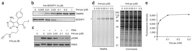
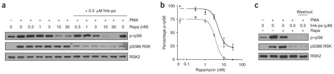
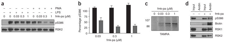
p90 ribosomal protein S6 kinases (RSKs) integrate upstream signals through two catalytic domains. Autophosphorylation of Ser386 by the regulatory C-terminal kinase domain (CTD) is thought to be essential for activation of the N-terminal kinase domain (NTD), which phosphorylates multiple downstream targets. We recently reported fmk, an irreversible inhibitor of the CTD of RSK1 and RSK2. Here we describe fmk-pa, a propargylamine variant that has improved cellular potency and a 'clickable' tag for assessing the extent and selectivity of covalent RSK modification. Copper-catalyzed conjugation of an azidoalkyl reporter (the click reaction) revealed that fmk-pa achieves selective and saturable modification of endogenous RSK1 and RSK2 in mammalian cells. Saturating concentrations of fmk-pa inhibited Ser386 phosphorylation and downstream signaling in response to phorbol ester stimulation, but had no effect on RSK activation by lipopolysaccharide. RSK autoactivation by the CTD is therefore context dependent, which suggests that NTD and CTD inhibitors should have distinct physiological effects.


Conflict of interest statement

COMPETING INTERESTS STATEMENT

The authors declare that they have no competing financial interests.

Figures

Figure 1
Figure 1
Mechanism of RSK activation. fmk is a selective inhibitor of the RSK CTD, which is thought to mediate autophosphorylation of Ser386 in the RSK hydrophobic motif (HM).
Figure 2
Figure 2
fmk-BODIPY irreversibly targets RSK but has only modest selectivity and potency in cells. (a) Chemical structure of fmk-BODIPY, a fluorescent fmk derivative. (b) Covalent labeling of RSK2 CTD by fmk-BODIPY. RSK2 CTD was treated with the indicated concentrations of unlabeled fmk for 1 h and then with 3 μM fmk-BODIPY for 1 h. Proteins were resolved by 10% SDS-PAGE and detected by in-gel fluorescence scanning, followed by western blot with an antibody to RSK2. (c) Effect of fmk-BODIPY on PMA-stimulated RSK Ser386 phosphorylation. HEK 293 cells were deprived of serum for 2 h and then treated with the indicated concentrations of fmk-BODIPY for 1 h. Cells were stimulated with PMA (0.1 μg ml−1) for 30 min and harvested in PBS. Proteins were resolved by 10% SDS-PAGE and detected by western blot with antibodies to phospho-Ser386 RSK (pS386) and total RSK2. (d) Selectivity of protein modification by fmk-BODIPY in intact cells. Proteins in cell lysates from c were resolved by 10% SDS-PAGE and detected by in-gel fluorescence scanning (left) and Coomassie blue staining (right). The asterisk marks the position of RSK1 and RSK2.
Figure 3
Figure 3
Irreversible, selective and potent inhibition of RSK by fmk-pa. (a) Chemical structure of fmk-pa, a clickable variant of fmk. (b) Click chemistry–mediated detection of RSK2 CTD. RSK2 CTD was treated with the indicated concentrations of fmk-pa for 1 h followed by 3 μM fmk-BODIPY for 1 h. After click conjugation with TAMRA-N3 (see Methods), proteins were resolved by 10% SDS-PAGE and detected by sequential in-gel fluorescence scanning using filters specific for TAMRA and BODIPY. (c) Effect of fmk-pa on PMA-stimulated Ser386 RSK phosphorylation. HEK 293 cells were deprived of serum for 2 h and then treated with the indicated concentrations of fmk-pa for 1 h. Cells were stimulated with PMA (0.1 μg ml−1) for 30 min and processed as described for Figure 2c. (d) Selectivity of protein modification by fmk-pa in intact cells. Cell lysates from c were subjected to click conjugation with TAMRA-N3. Proteins were resolved by 10% SDS-PAGE and detected by in-gel fluorescence scanning (left) and Coomassie blue staining (right). The asterisk marks the position of RSK1 and RSK2. (e) Quantification of fluorescent bands in d.
Figure 4
Figure 4
fmk-pa reveals a RSK CTD–dependent pathway for rpS6 phosphorylation. (a) Effect of rapamycin and fmk-pa on rpS6 phosphorylation. Serum-starved HEK 293 cells were treated with the indicated concentrations of rapamycin in the absence (left lanes) or presence (right lanes) of 300 nM fmk-pa for 1 h and stimulated with PMA (0.1 μg ml−1) for 30 min. Proteins were resolved by 10% SDS-PAGE and detected by western blot with antibodies to phospho-Ser235/236 rpS6 (p-rpS6), phospho-Ser386 RSK and total RSK2. (b) Dose-response curves showing additive inhibition of rpS6 phosphorylation: rapamycin alone, black triangles; rapamycin plus 300 nM fmk-pa, gray circles. Percentage rpS6 Ser235/236 phosphorylation was determined by quantification of western blots relative to DMSO controls using ImageJ (NIH). Data represent mean ± s.e.m. from three independent experiments. (c) Irreversible inhibition of rpS6 and RSK phosphorylation by fmk-pa. Serum-starved HEK 293 cells were treated with either fmk-pa (300 nM) or DMSO for 1 h. In the washout sample, fmk-pa was removed and cells were washed with compound-free medium and incubated with rapamycin (30 nM) for 1 h. Cells were stimulated with PMA (0.1 μg ml−1) for 30 min and processed as described for Figure 4a.
Figure 5
Figure 5
fmk-pa and click chemistry reveal a CTD-independent pathway for RSK Ser386 phosphorylation. (a) Effect of fmk-pa on Ser386 phosphorylation in primary bone marrow–derived macrophages stimulated with LPS or PMA. Macrophages were cultured in 0.5% serum for 20 h and then treated with the indicated concentrations of fmk-pa for 1 h. Cells were stimulated with either PMA (0.1 μg ml−1) or LPS (1 μg ml−1) for 30 min and harvested in PBS. Proteins were resolved by 10% SDS-PAGE and detected by western blot with antibodies to phospho-Ser386 RSK and total RSK2. (b) Percentage Ser386 phosphorylation was determined by quantification of western blots relative to DMSO controls (PMA-treated, gray bars; LPS-treated, black bars). Data represent mean ± s.e.m. from three independent experiments. (c) Saturable RSK modification by pretreatment of intact macrophages with fmk-pa. Lysates from LPS-stimulated macrophages (pretreated for 1 h with the indicated concentrations of fmk-pa) were incubated with TAMRA-N3 under the click reaction conditions. Proteins were resolved by 10% SDS-PAGE and detected by in-gel fluorescence scanning. (d) Use of click chemistry to demonstrate that fmk-pa–modified RSK1 and RSK2 are phosphorylated at Ser386 in response to LPS stimulation. Lysates from LPS-stimulated cells (pretreated with 1 μM fmk-pa or DMSO control) were incubated with biotin-N3 under the click reaction conditions for 1 h. Biotinylated proteins were affinity purified with avidin-agarose beads. Eluted proteins were resolved by 10% SDS-PAGE and detected by western blot with streptavidin-HRP or antibodies to RSK1, RSK2 and phospho-Ser386 RSK.

Comment in

References

    1. Hauge C, Frodin M. RSK and MSK in MAP kinase signalling. J Cell Sci. 2006;119:3021–3023. - PubMed
    1. Cohen MS, Zhang C, Shokat KM, Taunton J. Structural bioinformatics-based design of selective, irreversible kinase inhibitors. Science. 2005;308:1318–1321. - PMC - PubMed
    1. Smith JA, Poteet-Smith CE, Malarkey K, Sturgill TW. Identification of an extracellular signal-regulated kinase (ERK) docking site in ribosomal S6 kinase, a sequence critical for activation by ERK in vivo. J Biol Chem. 1999;274:2893–2898. - PubMed
    1. Gavin AC, Nebreda ARA. MAP kinase docking site is required for phosphorylation and activation of p90(rsk)/MAPKAP kinase-1. Curr Biol. 1999;9:281–284. - PubMed
    1. Frodin M, Jensen CJ, Merienne K, Gammeltoft S. A phosphoserine-regulated docking site in the protein kinase RSK2 that recruits and activates PDK1. EMBO J. 2000;19:2924–2934. - PMC - PubMed

Publication types

MeSH terms

LinkOut - more resources