. 2007 Feb 21;129(7):1959-67.
doi: 10.1021/ja065930i. Epub 2007 Jan 30.

DNA-encoded antibody libraries: a unified platform for multiplexed cell sorting and detection of genes and proteins

Affiliations

DNA-encoded antibody libraries: a unified platform for multiplexed cell sorting and detection of genes and proteins

Ryan C Bailey et al. J Am Chem Soc. .

Abstract

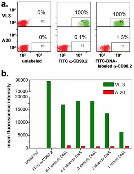
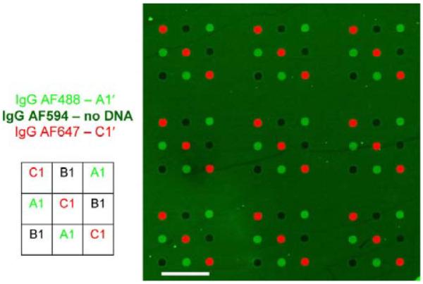
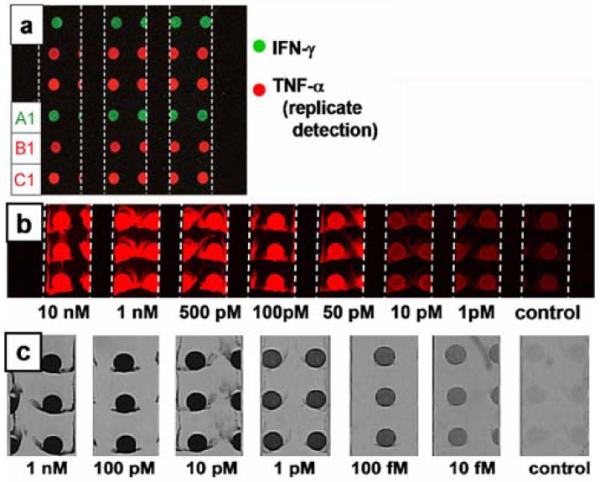
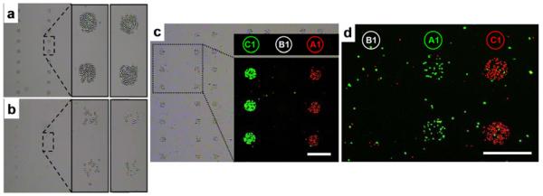
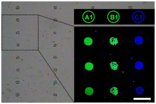
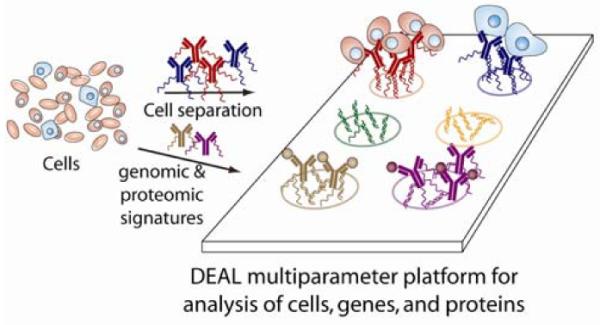
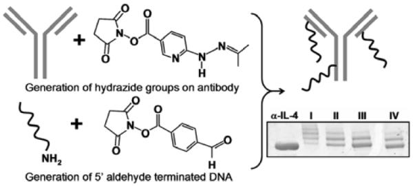
Whether for pathological examination or for fundamental biology studies, different classes of biomaterials and biomolecules are each measured from a different region of a typically heterogeneous tissue sample, thus introducing unavoidable sources of noise that are hard to quantitate. We describe the method of DNA-encoded antibody libraries (DEAL) for spatially multiplexed detection of ssDNAs and proteins as well as for cell sorting, all on the same diagnostic platform. DEAL is based upon the coupling of ssDNA oligomers onto antibodies which are then combined with the biological sample of interest. Spotted DNA arrays, which are found to inhibit biofouling, are utilized to spatially stratify the biomolecules or cells of interest. We demonstrate the DEAL technique for (1) the rapid detection of multiple proteins within a single microfluidic channel, and, with the additional step of electroless amplification of gold-nanoparticle labeled secondary antibodies, we establish a detection limit of 10 fM for the protein IL-2, 150 times more sensitive than the analogue ELISA; (2) the multiplexed, on-chip sorting of both immortalized cell lines and primary immune cells with an efficiency that exceeds surface-confined panning approaches; and (3) the co-detection of ssDNAs, proteins, and cell populations on the same platform.


Figures

Figure 1
Figure 1
Optimization of DNA loading of DEAL antibodies for cell surface marker recognition. (a). FACS plot comparing α-CD90.2/FITC-DNA conjugates with the commercially-available FITC α-CD90.2 antibody (no DNA). The conjugates bind to VL3 cells (100%) with minimal non-specific interactions with A20 (1.3%). When compared with FITC α-CD90.2, the overall fluorescent intensities are lower by a factor of 10, with slightly higher non-specific binding to A20. (b). Histogram of the mean fluorescent intensities for various FITC-DNA loadings. Fluorescence increases are roughly linear when the number of DNA strands is increased from 1 to 2 to 3, corresponding to the 1, 2 and 3 chromophores (1 per strand). For higher loadings, the fluorescence plateaus and then decreases.
Figure 2
Figure 2
Illustration of the resistance of the DEAL approach towards non-specific protein absorption. A microarray was simultaneously exposed to goat α-human IgG-Alexa488/A1', goat α-human IgGAlexa647/C1' DEAL conjugates and goat α-human IgG-Alexa594 with no pendant DNA. When the arrays were not fully blocked and/or rinsed, non-specific binding was observed on the surface of the glass slide, but not on the non-complementary spots of printed DNA, i.e., spot B1 did not have fluorescence from non-complementary IgG conjugates nor did it exhibit fluorescence from proteins not encoded with DNA (goat α-human IgGAlexa594). Scale bar corresponds to 1mm.
Figure 3
Figure 3
Fluorescence and brightfield images of DNA-templated protein immunoassays executed within microfluidic channels. The 600 μm micrometer wide channels are delineated with white dashed lines. (a). Two parameter DEAL immunoassay showing the detection of IFN-γ at spot A1 with a PE labeled 2° antibody (green channel) and replicate detection of TNF-α at spots B1 and C1 with an APC labeled 2° antibody (red channel). (b). Human IL-2 concentration series visualized using a fluorescent 2° antibody for detection. (c). Human IL-2 concentration series developed using Au electroless deposition as a visualization and amplification strategy.
Figure 4
Figure 4
The optimization and use of DEAL for multiplexed cell sorting. (a & b). Brightfield images showing the efficiency of the homogeneous DEAL cell capture process. (a). A homogeneous assay in which DEAL labeled antibodies are combined with the cells, and then the mixture is introduced onto the spotted DNA array microchip. (b). DEAL labeled antibodies are first assembled onto a spotted DNA array, followed by introduction of the cells. This heterogeneous process is similar to the traditional panning method of using surface bound antibodies to trap specific cells. The homogeneous process is clearly much more efficient. (c). Brightfield and fluorescence microscopy images of multiplexed cell sorting experiments where a 1:1 mixture of mRFP-expressing T cells (red channel) and EGFP-expressing B cells (green channel) is spatially stratified onto spots A1 and C1, corresponding to the encoding of α-CD90.2 and α-B220 antibodies with A1' and C1', respectively. (d). Fluorescence micrograph of multiplexed sorting of primary cells harvested from mice. A 1:1 mixture of CD4+ cells from EGFP transgenic mice and CD8+ cells from dsRed transgenic mice are separated to spots A1 and C1 by utilizing DEAL conjugates α-CD4-A1' and α-CD8-C1', respectively.
Figure 5
Figure 5
Microscopy images demonstrating simultaneous cell capture at spot B1 and multiparameter detection of genes and proteins, at spots A1 and C1, respectively. The brightfield image shows EGFP-expressing B cells (green channel) located to spots B1, FITC-labeled (green) cDNA at A1, and an APC-labeled TNF-α sandwich immunoassay (blue) encoded to C1. The scale bar corresponds to 300 μm.
Scheme 1
Scheme 1
Illustration of the DEAL method for cell sorting and co-detection of proteins and cDNAs (mRNAs). Antibodies against proteins (for cell sorting) or other proteins (including cell surface markers) are labeled with distinct DNA oligomers. These conjugates may then be combined with the biological sample (cells, tissue, etc.) where they bind to their cognate antigens. When introduced onto a DNA microarray, parallel self assembly, according to Watson-Crick base pairing, localizes the bound species to a specific spatial location allowing for multiplexed, multiparameter analysis.
Scheme 2
Scheme 2
Illustration of the two step coupling strategy utilized to prepare DEAL antibodies. In parallel, hydrazide groups are introduced onto a monoclonal antibody and 5' aldehyde modified single-stranded DNA is prepared from 5' aminated oligomers. When combined, hydrazone bonds are formed, linking the ssDNA to the antibody. At bottom right is a gel mobility shift assay showing varied oligomer (strand A1') loading unto α-human IL-4. By varying the stoichiometric ratios of SANH to antibody (lanes I–IV corresponds to 300:1, 100:1, 50:1, 25:1 respectively), the average number of attached oligonucleotides can be controlled.

References

    1. Lin B, White JT, Lu W, Xie T, Utleg AG, Yan X, Yi EC, Shannon P, Khretbukova I, Lange PH, Goodlett DR, Zhou D, Vasicek TJ, Hood L. Cancer Res. 2005;65:3081–3091. - PubMed
    1. Kwong KY, Bloom GC, Yang I, Boulware D, Coppola D, Haseman J, Chen E, McGrath A, Makusky AJ, Taylor J, Steiner S, Zhou J, Yeatman TJ, Quackenbush J. Genomics. 2005;86:142–158. - PubMed
    1. Huber M, Bahr I, Kratzchmar JR, Becker A, Muller E-C, Donner P, Pohlenz H-D, Schneider MR, Sommer A. Molec. Cell. Proteomics. 2004;3:43–55. - PubMed
    1. Tian Q, Stepaniants SB, Mao M, Weng L, Feetham MC, Doyle MJ, Yi EC, Dai H, Thorsson V, Eng J, Goodlett D, Berger JP, Gunter B, Linseley PS, Stoughton RB, Aebersold R, Collins SJ, Hanlon WA, Hood LE. Molec. Cell. Proteomics. 2004;3:960–969. - PubMed
    1. Chen G, Gharib TG, Huang C-C, Taylor JMG, Misek DE, Kardia SLR, Giordano TJ, Iannettoni MD, Orringer MB, Hanash SM, Beer DG. Molec. Cell. Proteomics. 2002;1:304–313. - PubMed

Publication types