Comparative Study
. 2007 Feb 20;46(7):1941-51.
doi: 10.1021/bi0622524. Epub 2007 Jan 30.

X-ray crystal structure of aristolochene synthase from Aspergillus terreus and evolution of templates for the cyclization of farnesyl diphosphate

Affiliations
Comparative Study

X-ray crystal structure of aristolochene synthase from Aspergillus terreus and evolution of templates for the cyclization of farnesyl diphosphate

Ekaterina Y Shishova et al. Biochemistry. .

Abstract

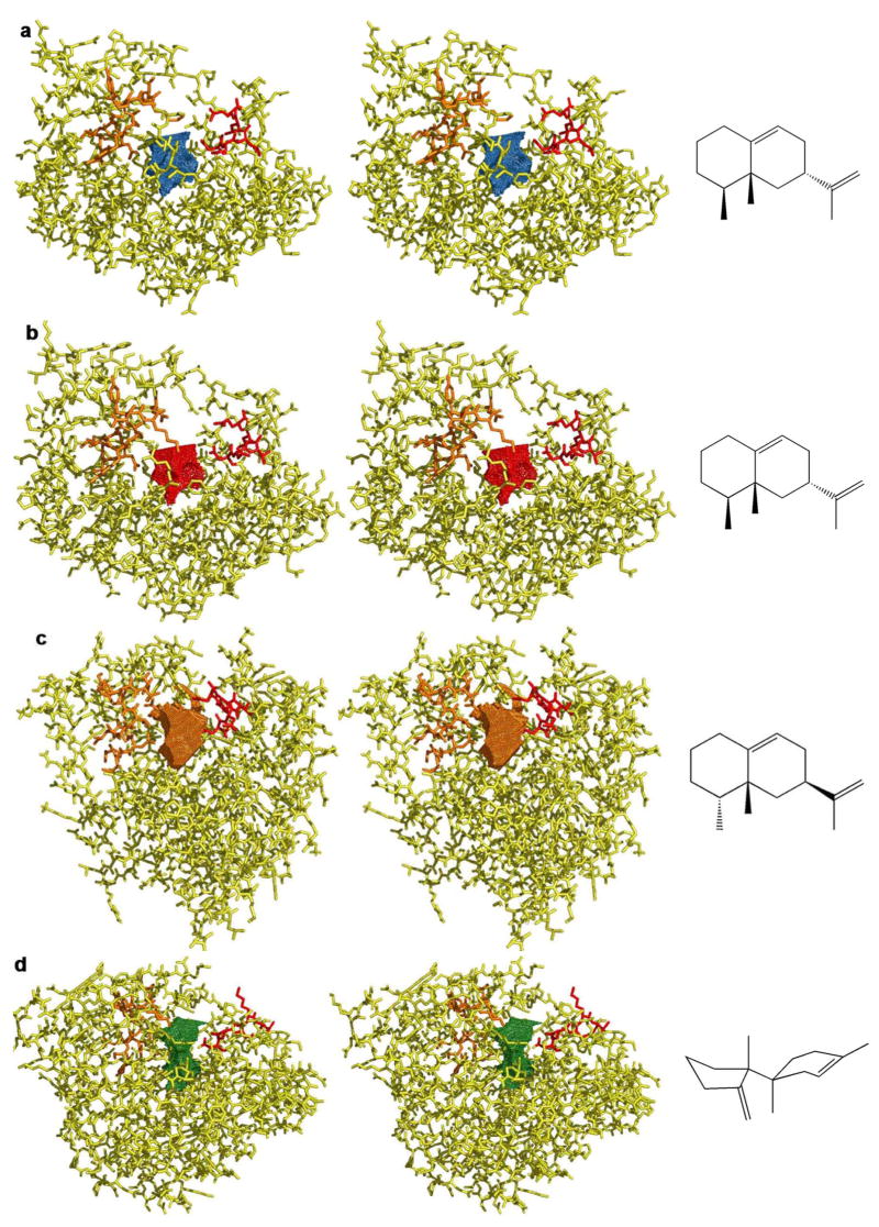
Aristolochene synthase from Aspergillus terreus catalyzes the cyclization of the universal sesquiterpene precursor, farnesyl diphosphate, to form the bicyclic hydrocarbon aristolochene. The 2.2 A resolution X-ray crystal structure of aristolochene synthase reveals a tetrameric quaternary structure in which each subunit adopts the alpha-helical class I terpene synthase fold with the active site in the "open", solvent-exposed conformation. Intriguingly, the 2.15 A resolution crystal structure of the complex with Mg2+3-pyrophosphate reveals ligand binding only to tetramer subunit D, which is stabilized in the "closed" conformation required for catalysis. Tetramer assembly may hinder conformational changes required for the transition from the inactive open conformation to the active closed conformation, thereby accounting for the attenuation of catalytic activity with an increase in enzyme concentration. In both conformations, but especially in the closed conformation, the active site contour is highly complementary in shape to that of aristolochene, and a catalytic function is proposed for the pyrophosphate anion based on its orientation with regard to the presumed binding mode of aristolochene. A similar active site contour is conserved in aristolochene synthase from Penicillium roqueforti despite the substantial divergent evolution of these two enzymes, while strikingly different active site contours are found in the sesquiterpene cyclases 5-epi-aristolochene synthase and trichodiene synthase. Thus, the terpenoid cyclase active site plays a critical role as a template in binding the flexible polyisoprenoid substrate in the proper conformation for catalysis. Across the greater family of terpenoid cyclases, this template is highly evolvable within a conserved alpha-helical fold for the synthesis of terpene natural products of diverse structure and stereochemistry.


Figures

Figure 1
Figure 1
Cyclization of farnesyl diphosphate (1) to form (+)-aristolochene (4). Mechanistic details are summarized in the text; the pyrophosphate leaving group of the substrate may serve as a general acid/general base in catalysis.
Figure 2
Figure 2
Ribbon plots of A. terreus aristolochene synthase. (a) Stereoview of cyclase monomer A showing the aspartate-rich motif (red) and the aspartate-poor NSE motif (orange) flanking the mouth of the active site. Helices are labeled according to the convention first established for farnesyl diphosphate synthase (28). (b) Cyclase monomers A (blue), B (green), C (purple), and D (grey) assemble to form a dimer of dimers. (c) Same as (b) but rotated by 90°.
Figure 2
Figure 2
Ribbon plots of A. terreus aristolochene synthase. (a) Stereoview of cyclase monomer A showing the aspartate-rich motif (red) and the aspartate-poor NSE motif (orange) flanking the mouth of the active site. Helices are labeled according to the convention first established for farnesyl diphosphate synthase (28). (b) Cyclase monomers A (blue), B (green), C (purple), and D (grey) assemble to form a dimer of dimers. (c) Same as (b) but rotated by 90°.
Figure 3
Figure 3
Native gel analysis of A. terreus aristolochene synthase oligomerization in solution. Novex Tris-glycine sample native buffer was added to 28 μM, 20 nM, and 11 mM enzyme in 25 mM MES (pH 6.5), 2 mM MgCl2, 0.15 M NaCl, 4 mM BME. The NativeMark Unstained protein standard from Invitrogen and enzyme samples were electrophoresed on 10–20% native Tris Glycine gel at 125 V and 4 °C in 50 mM Tris-glycine buffer (pH 8.8) for approximately 5 hours. Lanes labeled 1,2 and 3 indicate 28 μM, 20 nM, and 11 mM aristolochene synthase, respectively. The native enzyme migrates as a ~70 kDa dimer.
Figure 4
Figure 4
Aristolochene synthase–Mg2+3-PPi complex. (a) Simulated annealing omit maps of the PPi anion (blue) and Mg2+ ions (red) contoured at 3σ. The Mg2+ ions are shown as grey spheres and water molecules appear as red spheres. (b) Same orientation as (a) showing selected metal coordination and hydrogen bond interactions. (c) Superposition of aristolochene synthase and trichodiene synthase (green) active sites in their Mg2+3-PPi complexes. (d) Least-square superposition of 300 Cα atoms between unliganded (blue) and Mg 2+3-complexed (green) aristolochene synthase. Glycerol molecules are omitted from both structures for clarity. Significant conformational changes are triggered in the indicated helices and loops by Mg2+3-PPi binding.
Figure 5
Figure 5
(a) Superposition of aristolochene synthases from A. terreus (blue) and P. roqueforti (green). Aristolochene synthase from P. roqueforti crystallizes as a dimer along a two-fold noncrystallographic axis. With 1630 Å2buried surface area, the dimer interface is identical to those between the AD and BC monomer pairs in the A. terreus enzyme. (b) Stereoview of aristolochene synthase from A. terreus with sites of amino acid substitutions in the P. roqueforti enzyme colored yellow. Aspartate-rich and NSE motifs are red and orange, respectively.
Figure 6
Figure 6
Left: stereoviews of sesquiterpene cyclases and their solvent accessible active site surface contours in the open, unliganded conformations: (a) aristolochene synthase from A. terreus, (b) aristolochene synthase from P. roqueforti, (c) C-terminal domain of 5-epi aristolochene synthase, (d) trichodiene synthase. Aspartate-rich motifs (red) and NSE motifs (orange) indicate the comparable orientations of active site clefts, roughly comparable to that shown in Figure 2a for aristolochene synthase from A. terreus. Right: cyclic sesquiterpenes generated by each enzyme.
Figure 7
Figure 7
Active site surface contours of A. terreus aristolochene synthase in the unliganded (from Figure 6a) and liganded (closed) conformation. The cyclization product, (+)-aristolochene, is modeled into the active site of the closed conformation, and the location of the Mg2+3-PPi cluster is shown as a visual reference. The unique template required for the cyclization of farnesyl diphosphate is preserved in the lower active site contour in both open and closed conformations. The O6 atom of PPi corresponds to the original phosphoester oxygen and the O3 atom corresponds to the terminal phosphate group of the substrate, farnesyl diphosphate. The proximity of the PPi O3 atom to aristolochene C6 and C8 atoms (3.4 Å and 3.2 Å, respectively) implicates PPi as the general acid/general base illustrated in Figure 1.

Similar articles

Cited by

References

    1. Glasby JS. Encyclopedia of Terpenoids. Wiley; Chichester: 1982.
    1. Pichersky E, Noel JP, Dudareva N. Biosynthesis of plant volatiles: Nature’s diversity and ingenuity. Science. 2006;311:808–811. - PMC - PubMed
    1. Cane DE. Isoprenoid biosynthesis. Stereochemistry of the cyclization of allylic pyrophosphates. Acc Chem Res. 1985;18:220–226.
    1. Cane DE. Enzymatic formation of sesquiterpenes. Chem Rev. 1990;90:1089–1103.
    1. Lesburg CA, Caruthers JM, Paschall CM, Christianson DW. Managing and manipulating carbocations in biology: Terpenoid cyclase structure and mechanism. Curr Opin Struct Biol. 1998;8:695–703. - PubMed

Publication types