

Distinct developmental changes in the distribution of calcium, phosphorus and sulphur during fetal growth-plate development
- PMID: 17261139
- PMCID: PMC2100269
- DOI: 10.1111/j.1469-7580.2006.00680.x
Distinct developmental changes in the distribution of calcium, phosphorus and sulphur during fetal growth-plate development
Abstract
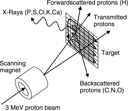
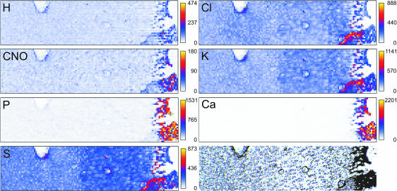
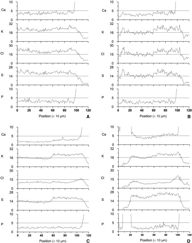
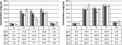
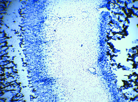
Gradients in the concentrations of free phosphate (Pi) and calcium (Ca) exist in fully developed growth zones of long bones and ribs, with the highest concentrations closest to the site of mineralization. As high concentrations of Pi and Ca induce chondrocyte maturation and apoptosis, it has been hypothesized that Ca and Pi drive chondrocyte differentiation in growth plates. This study aimed to determine whether gradients in the important spectral elements phosphorus (P), Ca and sulphur (S) are already present in early stages of development, or whether they gradually develop with maturation of the growth zone. We quantified the concentration profiles of Ca, P, S, chloride and potassium at four different stages of early development of the distal growth plates of the porcine femurs, using particle-induced X-ray emission and forward- and backward-scattering spectrometry with a nuclear microprobe. A Ca concentration gradient towards the mineralized area and a stepwise increase in S was found to develop slowly with tissue maturation. The increase in S co-localizes with the onset of proliferation. A P gradient was not detected in the earliest developmental stages. High Ca levels, which may induce chondrocyte maturation, are present near the mineralization front. As total P concentrations do not correspond with former free Pi measurements, we hypothesize that the increase of free Pi towards the bone-forming site results from enzymatic cleavage of bound phosphate.
Figures
References
-
- Alini M, Carey D, Hirata S, Grynpas MD, Pidoux I, Poole AR. Cellular and matrix changes before and at the time of calcification in the growth plate studied in vitro: arrest of type X collagen synthesis and net loss of collagen when calcification is initiated. J Bone Miner Res. 1994;9:1077–1087. - PubMed
-
- Brands PJM, Mutsaers PHA, De Voigt MJA. System for on-line monitoring of light element concentration distributions in thin samples. Nuclear Instruments and Methods in Physics Research, Section B: Beam Interactions with Materials and Atoms. 1999;158:135–140.
-
- Burton DW, Foster M, Johnson KA, Hiramoto M, Deftos LJ, Terkeltaub R. Chondrocyte calcium-sensing receptor expression is up-regulated in early guinea pig knee osteoarthritis and modulates PTHrP, MMP-13, and TIMP-3 expression. Osteoarthritis Cartilage. 2005;13:395–404. - PubMed
-
- Byers S, van Rooden JC, Foster BK. Structural changes in the large proteoglycan, aggrecan, in different zones of the ovine growth plate. Calcified Tissue Int. 1997;60:71–78. - PubMed
MeSH terms
Substances
LinkOut - more resources
Full Text Sources
Research Materials
Miscellaneous
