

Comparison of manual and semi-automated delineation of regions of interest for radioligand PET imaging analysis
- PMID: 17261193
- PMCID: PMC1802071
- DOI: 10.1186/1471-2385-7-2
Comparison of manual and semi-automated delineation of regions of interest for radioligand PET imaging analysis
Abstract
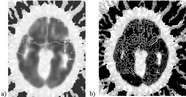
Background: As imaging centers produce higher resolution research scans, the number of man-hours required to process regional data has become a major concern. Comparison of automated vs. manual methodology has not been reported for functional imaging. We explored validation of using automation to delineate regions of interest on positron emission tomography (PET) scans. The purpose of this study was to ascertain improvements in image processing time and reproducibility of a semi-automated brain region extraction (SABRE) method over manual delineation of regions of interest (ROIs).
Methods: We compared 2 sets of partial volume corrected serotonin 1a receptor binding potentials (BPs) resulting from manual vs. semi-automated methods. BPs were obtained from subjects meeting consensus criteria for frontotemporal degeneration and from age- and gender-matched healthy controls. Two trained raters provided each set of data to conduct comparisons of inter-rater mean image processing time, rank order of BPs for 9 PET scans, intra- and inter-rater intraclass correlation coefficients (ICC), repeatability coefficients (RC), percentages of the average parameter value (RM%), and effect sizes of either method.
Results: SABRE saved approximately 3 hours of processing time per PET subject over manual delineation (p < .001). Quality of the SABRE BP results was preserved relative to the rank order of subjects by manual methods. Intra- and inter-rater ICC were high (>0.8) for both methods. RC and RM% were lower for the manual method across all ROIs, indicating less intra-rater variance across PET subjects' BPs.
Conclusion: SABRE demonstrated significant time savings and no significant difference in reproducibility over manual methods, justifying the use of SABRE in serotonin 1a receptor radioligand PET imaging analysis. This implies that semi-automated ROI delineation is a valid methodology for future PET imaging analysis.
Figures
Similar articles
-
Inter-rater reliability of manual and automated region-of-interest delineation for PiB PET.Neuroimage. 2011 Apr 1;55(3):933-41. doi: 10.1016/j.neuroimage.2010.12.070. Epub 2010 Dec 31. Neuroimage. 2011. PMID: 21195782 Free PMC article.
-
An automated method for the extraction of regional data from PET images.Psychiatry Res. 2006 Jun 30;147(1):79-89. doi: 10.1016/j.pscychresns.2006.01.011. Epub 2006 Jun 21. Psychiatry Res. 2006. PMID: 16797168
-
Evaluation of two automated methods for PET region of interest analysis.Neuroinformatics. 2014 Oct;12(4):551-62. doi: 10.1007/s12021-014-9233-6. Neuroinformatics. 2014. PMID: 24880728
-
Performance Evaluation of a Semi-automated Method for [18F]FDG Uptake in Abdominal Visceral Adipose Tissue.Mol Imaging Biol. 2019 Feb;21(1):159-167. doi: 10.1007/s11307-018-1211-1. Mol Imaging Biol. 2019. PMID: 29789994
-
A Manual of the Nervous Diseases of Man.Br Foreign Med Rev. 1844 Apr;17(34):407-423. Br Foreign Med Rev. 1844. PMID: 30162178 Free PMC article. Review. No abstract available.
Cited by
-
Lesion Explorer: a video-guided, standardized protocol for accurate and reliable MRI-derived volumetrics in Alzheimer's disease and normal elderly.J Vis Exp. 2014 Apr 14;(86):50887. doi: 10.3791/50887. J Vis Exp. 2014. PMID: 24797507 Free PMC article.
-
[11C]SCH23390 binding to the D1-dopamine receptor in the human brain-a comparison of manual and automated methods for image analysis.EJNMMI Res. 2018 Aug 2;8(1):74. doi: 10.1186/s13550-018-0416-2. EJNMMI Res. 2018. PMID: 30069645 Free PMC article.
-
Artificial intelligence in molecular imaging.Ann Transl Med. 2021 May;9(9):824. doi: 10.21037/atm-20-6191. Ann Transl Med. 2021. PMID: 34268437 Free PMC article. Review.
References
-
- Dade LA, Gao FQ, Kovacevic N, Roy P, Rockel C, O'Toole CM, Lobaugh NJ, Feinstein A, Levine B, Black SE. Semiautomatic brain region extraction: A method of parcellating brain regions from structural magnetic resonance images. Neuroimage. 2004;22:1492–1502. doi: 10.1016/j.neuroimage.2004.03.023. - DOI - PubMed
-
- Caviness Jr VS, Meyer J, Makris N, Kennedy DN. MRI-based topographic parcellation of human neocortex: An anatomically specified method with estimate of reliability. Journal of Cognitive Neuroscience. 1996;8:566–587. - PubMed
-
- Chan D, Fox NC, Jenkins R, Scahill RI, Crum WR, Rossor MN. Rates of global and regional cerebral atrophy in AD and frontotemporal dementia. Neurology. 2001;27:1756–1763. - PubMed
Grants and funding
LinkOut - more resources
Full Text Sources
Research Materials
