. 2007 Apr;27(7):2512-26.
doi: 10.1128/MCB.01907-06. Epub 2007 Jan 29.

Dss1 interaction with Brh2 as a regulatory mechanism for recombinational repair

Affiliations

Dss1 interaction with Brh2 as a regulatory mechanism for recombinational repair

Qingwen Zhou et al. Mol Cell Biol. 2007 Apr.

Abstract

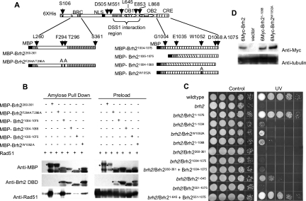
Brh2, the BRCA2 ortholog in Ustilago maydis, enables recombinational repair of DNA by controlling Rad51 and is in turn regulated by Dss1. Interplay with Rad51 is conducted via the BRC element located in the N-terminal region of the protein and through an unrelated domain, CRE, at the C terminus. Mutation in either BRC or CRE severely reduces functional activity, but repair deficiency of the brh2 mutant can be complemented by expressing BRC and CRE on different molecules. This intermolecular complementation is dependent upon the presence of Dss1. Brh2 molecules associate through the region overlapping with the Dss1-interacting domain to form at least dimer-sized complexes, which in turn, can be dissociated by Dss1 to monomer. We propose that cooperation between BRC and CRE domains and the Dss1-provoked dissociation of Brh2 complexes are requisite features of Brh2's molecular mechanism.


Figures

FIG. 1.
FIG. 1.
Mapping the C-terminal Rad51-interacting region, CRE. (A) Brh2 is illustrated schematically with the BRC domain, helical domain, OB1 and OB2 with the inserted tower, and the CRE region regions represented. The boundaries of the truncation mutations or the particular point mutations are shown. The allele product representing completely functional Brh2 started at residue 106 as discussed in Materials and Methods. (B) The indicated fragments were expressed as MBP fusions in E. coli in the combinations shown in the matrix. Pull-down assays were performed with cleared cell extracts using amylose beads to capture MBP-tagged proteins. After specific elution of associated proteins from the beads by using maltose-containing buffer, samples were electophoresed in 10% polyacrylamide gels with sodium dodecyl sulfate and analyzed by Western blotting. MBP-Brh2 fusions are distinguished with asterisks. (C) Survival of brh2 mutant cells expressing the indicated Brh2 mutant allele products. Serial 10-fold dilutions of cell suspensions were spotted from left to right and plates were irradiated with 120 J/m2 UV light. (D) Western blot analysis of 6Myc-tagged Brh2 alleles. Extracts of UCM565 cells expressing the indicated allele products were immunoprecipitated with anti-myc antibodies and protein A agarose. Bound protein was electrophoresed in an SDS-polyacrylamide gel, transferred to membrane, and probed with anti-myc monoclonal antibody to detect tagged Brh2. A loading control using the cell extracts was performed with anti-β-tubulin antibodies.
FIG. 2.
FIG. 2.
Determinants of Brh2 association. (A) MBP- and His-Brh2 fusions were coexpressed in the presence or absence of Rad51, and pull-down assays were performed with amylose beads used to capture MBP-Brh2 and associated proteins. The allele product representing completely functional Brh2 started at residue 106 as discussed in Materials and Methods. (B) Aliquots of cell extracts containing 1 mM MgCl2 were pretreated by incubation with 2 μg per ml DNase I and RNase A at 4°C for 12 h before pull-down assays were performed.
FIG. 3.
FIG. 3.
Pull-down mapping of Brh2 interaction domains. (A) Schematic illustration of MBP-tagged Brh2 fragments used in pull-down assays with His-tagged Brh2 or an internal fragment. A summary of the pull-down results below is presented schematically on the right with pluses and minuses to indicate the relative strengths of association (affinities). The data for interaction of the full-length MBP-Brh2 with His-Brh2 are presented in Fig. 2B. (B) Pull-down assays were performed with Ni-NTA resin to capture the His-tagged bait protein, either His-Brh2 (Brh2106-1075) or His-Brh2505-853. After the resin processing and elution with 200 mM imidazole, Western blotting was performed using anti-His monoclonal antibody and anti-MBP antiserum. Since the MBP-Brh2 allele products being tested were of different lengths, their mobilities during SDS gel electrophoresis differed. To keep the figure compact and to allow for easy comparison of the levels of protein, lanes with different allele products were spliced from the same autoradiographic film as indicated by the boxes, and the boxes with the bands representing the Brh2 allele products were aligned (indicated as “MBP-Brh2 allele”). It is to be emphasized that the sizes of the Brh2 allele products are different from box to box, even though the bands are aligned as indicated. The sizes of the polypeptides are indicated by the amino acid residues of the MBP-tagged Brh2 allele product. The mapping results presented for the individual N-terminal, C-terminal, or medial regions came from the same autoradiographs under the same exposure. Background intensity varied somewhat due to the position of the bands relative to each other in the autoradiographs.
FIG. 4.
FIG. 4.
Gel filtration analysis of Brh2. Complexes of MBP-Brh2505-1075, His-Brh2505-1075 and His-Dss1 were affinity purified on Ni2+-NTA resin. (A) Complexes eluted from Ni2+-NTA with imidazole buffer containing 0.2 M KCl were chromatographed on Mono Q beads with the KCl gradient as shown. Fractions spanning the peak were collected as shown by the corresponding hatch marks underneath the trace and examined by SDS gel electrophoresis. Relative molecular masses of MBP-Brh2505-1075, His-Brh2505-1075 and His-Dss1 were approximately 100, 60, and 27 kDa, respectively. (B) Fraction 2 from the Mono Q peak was applied to a column of cross-linked amylose. The bound fraction was eluted with 10 mM maltose and then applied to a Superose 6 column in 0.2 M KCl, calibrated with the size markers thyroglobulin (660 kDa), ferritin (440 kDa), catalase (232 kDa), bovine serum albumin (67 kDa), ovalbumin (43 kDa), and RNase A (14 kDa). Fractions were collected as shown by the corresponding hatch marks underneath the trace, and molecular species were identified by Western blot analysis probing with anti-MBP antiserum and anti-His monoclonal antibody and by directly visualizing His-Dss1 by soaking the gels with InVision stain. (C) The flowthrough from the amylose column was applied to Superose 6 as described above. Fractions were analyzed by Western blotting to detect MBP-Brh2505-1075, His-Brh2505-1075, and His-Dss1. (D) Complexes eluted from Ni2+-NTA with imidazole containing 0.5 M NaCl were chromatographed on Mono Q beads as described above. Fractions spanning the heterogeneous mixture were collected as shown by the corresponding hatch marks and aliquots examined by SDS gel electrophoresis. Fractions 3 and 4 were pooled and applied to amylose resin. The fraction that bound to amylose (E) and the flowthrough (F) were analyzed by gel filtration as described above. MBP-Brh2505-1075, His-Brh2505-1075, and His-Dss1 peaked at fractions 35, 39, and 43, respectively. Fraction 27 corresponded to an approximately dimer-sized complex, while fraction 17 was larger than a tetramer. Fraction 7 corresponded to the excluded fraction.
FIG. 5.
FIG. 5.
Chemical cross-linking of Brh2. (A) His-Rad51, MBP-Brh2/His-Dss1, and ovalbumin at 2.5 μM were held in a reaction mixture containing BS3 as described in Materials and Methods. For His-Rad51 and MBP-Brh2/His-Dss1, reaction mixtures contained no reagent or contained BS3 in decreasing concentrations of 50 μM, 10 μM, and 2 μM, respectively. For ovalbumin, the reaction mixture contained 50 μM and 10 μM BS3 only. After the reactions, proteins were separated on an SDS gel with a 5 to 16% gradient of acrylamide. The asterisks indicate bands with the mobility of the expected dimeric form. (B) A parallel gel was cut into two at the position of the 50-kDa marker. The top half was transferred to a PVDF membrane and Western blotted using anti-His monoclonal antibody (top panel). After blotting, the membrane was dipped into Coomassie blue (CB) staining solution for 1 s and then immediately flushed with water and acetic acid/methanol destaining solution to visualize the electrophoretically transferred proteins. The bottom half of the gel was soaked in Invision stain to visualize the His-tagged proteins. The asterisks indicate bands with the mobility of the expected dimeric form.
FIG. 6.
FIG. 6.
BRC and CRE communication in trans depends upon the maintenance of plasmids expressing each. (A) Schematic illustration of Brh2 allele products with deletions of the BRC or CRE region. (B) Survival of brh2 mutant cells expressing the indicated Brh2 allele products after irradiation with 120 J/m2 UV light is shown. Brh2316-1075 (Brh2ΔBRC) was expressed from a plasmid with a HygR marker, while the Brh21-1034 (Brh2ΔCRE) plasmid carried a CbxR marker. Two independent clones of cells cured of one plasmid or the other were tested. (C) For selective loss of Brh2ΔBRC or Brh2ΔCRE measured in individual clones, cells cotransformed with both plasmids were cured of one plasmid or the other by propagation in liquid medium containing either hygromycin or carboxin. After being streaked for isolation on solid medium, 52 single colonies were picked and tested for loss of the unselected marker by sequentially patching onto plates with neither drug or one of the drugs. Across the top of each plate from left to right were three controls, brh2, wild type, and brh2 cotransformant, and then the 52 individuals. Two independent clones that retained one of the parental plasmids but that was cured of the other plasmid were then tested (see panel B) for survival after UV treatment. (D) Population-level loss of plasmid expressing Brh2ΔBRC or Brh2ΔCRE. Three independent cotransformants expressing both Brh2ΔBRC (HygR) and Brh2ΔCRE (CbxR) were cultured in liquid medium containing both drugs or one of the two drugs. After growth for 20 generations, aliquots were removed and diluted in a 10-fold serial dilutions, and then samples were plated on solid medium containing the indicated drug or else were irradiated with UV (120 J/m2).
FIG. 7.
FIG. 7.
BRC and CRE communication mediated by Dss1. (A) Survival of brh2 and dss1 single- or double-mutant cells at a representative UV dose of 20 J/m2. (B, top panel) GFP-Brh2 focus formation 2 h after irradiation with UV (30 J/m2). Cells with representative foci (see arrows) are shown. (B, bottom panel) Following irradiation, aliquots were removed at the indicated times and the percentage of cells with foci was determined after counting approximately 200 cells for each point. wt, wild type. (C) Survival after irradiation with 120 J/m2 UV light of brh2 dss1 double-mutant cells expressing the indicated BRC- and/or CRE-defective Brh2 allele products (Brh2316-1075 and Brh21-1034, respectively, as in Fig. 6) with and without Dss1.
FIG. 8.
FIG. 8.
Brh2 point mutant unresponsive to Dss1. (A) Schematic illustration of Brh2 point mutants and truncations. (B) Survival of brh2 mutant cells expressing the indicated alleles after irradiation with 120 J/m2 UV light. (C) MBP- and His-Brh2 fusions of Brh2W728 and Brh2106-1075 (wt) were coexpressed in E. coli in the presence or absence of His-Dss1, and pull-down assays were performed with amylose beads. Western analysis was performed as described for Fig. 2.
FIG. 9.
FIG. 9.
Dss1 switch model for regulating Brh2. Brh2 shown at the top in light gray has BRC and CRE Rad51-interacting domains located at N and C termini (unspecified). In the middle is the Brh2 self-interacting domain. Brh2 can associate with itself via the medial region and with Rad51 (black helmet shape) through cooperation between BRC and CRE domains in trans. Dss1 (dark banana shape) binds to a domain that overlaps with the Brh2 self-interacting region, promoting dissociation of Brh2 and concomitant presentation of Rad51 in a state suitable for initiating filament formation. No information on equilibrium constants or on the disposition of DNA is known.

Similar articles

Cited by

References

    1. Baillat, D., M. A. Hakimi, A. M. Naar, A. Shilatifard, N. Cooch, and R. Shiekhattar. 2005. Integrator, a multiprotein mediator of small nuclear RNA processing, associates with the C-terminal repeat of RNA polymerase II. Cell 123:631-640. - PubMed
    1. Chen, C. F., P. L. Chen, Q. Zhong, Z. D. Sharp, and W. H. Lee. 1999. Expression of BRC repeats in breast cancer cells disrupts the BRCA2-Rad51 complex and leads to radiation hypersensitivity and loss of G(2)/M checkpoint control. J. Biol. Chem. 274:32931-32935. - PubMed
    1. Clontech Laboratories. 2001. Yeast protocols handbook. PT3024-1. Clontech Laboratories, Palo Alto, CA.
    1. Davies, A. A., J. Y. Masson, M. J. McIlwraith, A. Z. Stasiak, A. Stasiak, A. R. Venkitaraman, and S. C. West. 2001. Role of BRCA2 in control of the RAD51 recombination and DNA repair protein. Mol. Cell 7:273-282. - PubMed
    1. Esashi, F., N. Christ, J. Gannon, Y. Liu, T. Hunt, M. Jasin, and S. C. West. 2005. CDK-dependent phosphorylation of BRCA2 as a regulatory mechanism for recombinational repair. Nature 434:598-604. - PubMed

Publication types