. 2007 Apr;75(4):1861-9.
doi: 10.1128/IAI.01473-06. Epub 2007 Jan 29.

Extracellular gelatinase of Enterococcus faecalis destroys a defense system in insect hemolymph and human serum

Affiliations

Extracellular gelatinase of Enterococcus faecalis destroys a defense system in insect hemolymph and human serum

Shin Yong Park et al. Infect Immun. 2007 Apr.

Abstract

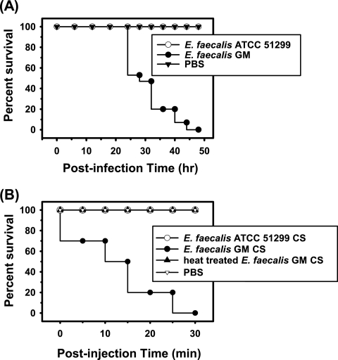
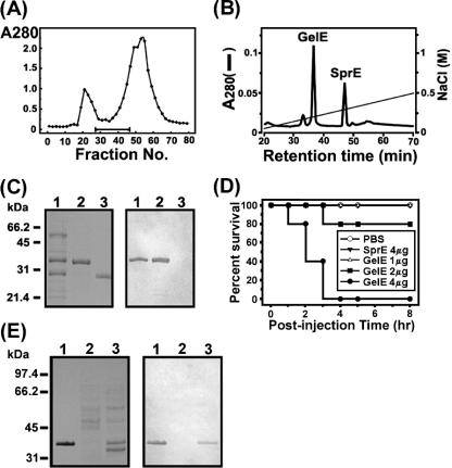
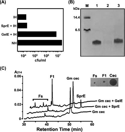
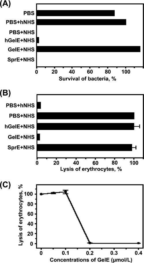
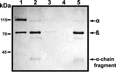
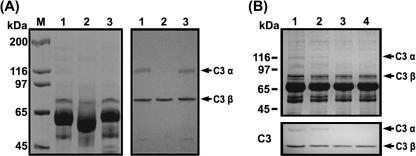
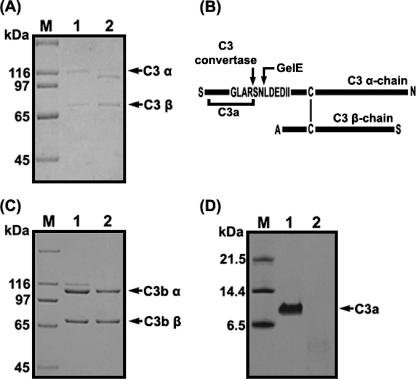
We isolated Enterococcus faecalis from the body fluids of dead larvae of the greater wax moth, Galleria mellonella. Extracellular gelatinase (GelE) and serine protease (SprE), both of which are considered putative virulence factors of E. faecalis, were purified from the culture supernatant of E. faecalis. In an attempt to elucidate their virulence mechanisms, purified GelE and SprE were injected into hemolymph of G. mellonella and evaluated with regard to their effects on the immune system of insect hemolymph. As a result, it was determined that E. faecalis GelE degraded an inducible antimicrobial peptide (Gm cecropin) which is known to perform a critical role in host defense during the early phase of microbial infection. The results obtained from the G. mellonella-E. faecalis infection model compelled us to assess the virulence activity of GelE against the complement system in human serum. E. faecalis GelE hydrolyzed C3a and also mediated the degradation of the alpha chain of C3b, thereby inhibiting opsonization and the formation of the membrane attack complex resultant from the activation of the complement cascade triggered by C3 activation. In contrast, E. faecalis SprE exhibited no virulence effect against the immune system of insect hemolymph or human serum tested in this study.


Figures

FIG. 1.
FIG. 1.
Survival of insects upon injection with E. faecalis GM or CS. Kaplan-Meier survival plots of G. mellonella injected with E. faecalis GM or ATCC 51299 (A) and CS of each strain (B). For a control, 5 μl of PBS was injected into each of the test larvae.
FIG. 2.
FIG. 2.
Purification of GelE from concentrated CS of E. faecalis GM. (A) Profile of Sephadex G-100 gel filtration chromatography. The optical density of each fraction was measured at 280 nm. The bar represents the range of fractions evidencing 100% mortality in our “infection and survival” test. (B) Anion-exchange chromatography as a final step in the purification of GelE. Two peaks, corresponding to gelatinase and serine protease, were indicated by GelE and SprE, respectively. (C) Tricine SDS-PAGE and immunoblot analysis. Left panel, a tricine SDS-PAGE gel stained with Coomassie blue; right panel, immunoblot analysis conducted with antiserum against GelE. Lane 1, pooled fractions (no. 26 to 46) from gel filtration chromatography, as indicated by the horizontal bar in panel A; lane 2, purified GelE; lane 3, purified SprE. (D) Kaplan-Meier survival plots of G. mellonella injected with differing quantities of purified GelE, ranging from 1 to 4 μg per larva, or SprE. Also, 5 μl of PBS was injected into the larvae as a control. (E) Detection of GelE in the concentrated CS of E. faecalis GM. Left panel, a 10% SDS-PAGE gel stained with Coomassie blue; right panel, immunoblot analysis using antiserum against GelE as a probe. Lane 1, purified GelE; lane 2, CS of E. faecalis ATCC 51299; lane 3, CS of E. faecalis GM.
FIG. 3.
FIG. 3.
Effects of GelE on antibacterial activity of G. mellonella hemolymph. (A) As shown in the acid extract sample of the immunized hemolymph incubated with GelE (GelE + IH), its antibacterial activity was significantly attenuated and close to that of nonimmunized hemolymph (NH). Error bars were too small to express. (B) A Coomassie blue-stained 16.5% tricine SDS-PAGE gel containing purified Gm cecropin and Gm cecropin treated with GelE or SprE. Lane M, standard marker; lane 1, 2 μg of Gm cecropin; lane 2, 2 μg of Gm cecropin treated with GelE; lane 3, 2 μg of Gm cecropin treated with SprE. (C) HPLC chromatograms of the GelE-induced digestion of Gm cecropin. Inset, radial diffusion assay for antibacterial activities of Fs, F1, and intact Gm cecropin (Cec).
FIG. 4.
FIG. 4.
Effect of GelE on complement activation in human seurm. (A) Role of GelE in complement-mediated killing of E. faecalis GM. The survival rate of E. faecalis GM in the PBS-hNHS sample was determined to be 100%, and the relative survival rate of each sample was measured. Error bars were too small to express. (B) Inhibitory effect of GelE on the complement-mediated lysis of rabbit erythrocytes. Lysis of erythrocytes in the PBS-NHS sample was determined to be 100%. (C) Dose-dependent effect of GelE on complement-mediated erythrocyte lysis. E. faecalis GM SprE was used as a negative control and had no effect on the complement-mediated lysis of erythrocytes (data not shown). Spontaneous erythrocyte lysis was <1% and was not represented.
FIG. 5.
FIG. 5.
Effect of GelE on cell surface deposition of C3. The protein samples on E. faecalis GM were detected via immunoblot analysis. The nitrocellulose membrane was probed with anti-C3 antibody, which had been diluted to a concentration of 1:1,000 in antibody solution (1% skim milk in Tris-buffered saline containing 0.05% Tween 20). The secondary antibody used in this experiment was horseradish peroxidase-conjugated goat anti-rabbit antibody, as appropriate, at a dilution of 1:8,000. One microgram of purified C3 was employed as a control. Lane 1, purified C3; lane 2, NHS incubated with PBS; lane 3, hNHS incubated with PBS; lane 4, NHS incubated with GelE; lane 5, NHS incubated with SprE. Arrows indicate the α chain, β chain, and α-chain fragments of human C3.
FIG. 6.
FIG. 6.
Degradation of complement factor C3 α chain by GelE, as determined via SDS-PAGE. (A) Proteolysis of the C3 α chain in NHS by GelE. Left panel, 8% SDS-PAGE gel conducted with NHS; right panel, immunoblot analysis. Lane M, molecular mass marker; lane 1, NHS incubated with PBS; lane 2, NHS incubated with GelE; lane 3, NHS incubated with SprE. Arrows indicate the intact α and β chains of C3 present in NHS. (B) Change in the C3 α chain in NHS by E. faecalis GM. Equal volumes of the mixtures were removed and subjected to SDS-PAGE and immunoblot analysis. Upper panel, an SDS-PAGE gel; lower panel, immunoblot analysis. Lane 1, NHS incubated with PBS; lane 2, NHS incubated with E. faecalis GM for 6 h; lane 3, NHS incubated with E. faecalis GM for 12 h; lane 4, NHS incubated with E. faecalis GM for 24 h.
FIG. 7.
FIG. 7.
Proteolysis of complement factor C3 and C3a by GelE. (A) SDS-PAGE performed with purified C3. Lane M, molecular mass marker; lane 1, C3 incubated with PBS; lane 2, C3 incubated with GelE. (B) Schematic view of C3 cleavage by GelE. The cleavage sites of C3 α chain by C3 convertase or GelE are marked by arrows, and the vertical bar represents a disulfide bond between the C3 α chain and C3 β chain. (C) SDS-PAGE performed with purified C3b. Lane M, molecular mass marker; lane 1, C3b incubated with PBS; lane 2, C3b incubated with GelE. (D) Tricine SDS-PAGE performed with purified C3a. Lane M, molecular mass marker; lane 1, C3a incubated with PBS; lane 2, C3a incubated with GelE. The gels were stained with Coomassie blue.

Similar articles

Cited by

References

    1. Akesson, P., A. G. Sjoholm, and L. Bjorck. 1996. Protein SIC, a novel extracellular protein of Streptococcus pyogenes interfering with complement function. J. Biol. Chem. 271:1081-1088. - PubMed
    1. Azghani, A. O., L. D. Gray, and A. R. Johnson. 1993. A bacterial protease perturbs the paracellular barrier function of transporting epithelial monolayers in culture. Infect. Immun. 61:2681-2686. - PMC - PubMed
    1. Baldassarri, L., R. Creti, L. Montanaro, G. Orefici, and C. R. Arciola. 2005. Pathogenesis of implant infections by enterococci. Int. J. Artif. Organs 28:1101-1109. - PubMed
    1. Choi, J. Y., C. D. Sifri, B. C. Goumnerov, L. G. Rahme, F. M. Ausubel, and S. B. Calderwood. 2002. Identification of virulence genes in a pathogenic strain of Pseudomonas aeruginosa by representational difference analysis. J. Bacteriol. 184:952-961. - PMC - PubMed
    1. Coburn, P. S., and M. S. Gilmore. 2003. The Enterococcus faecalis cytolysin: a novel toxin active against eukaryotic and prokaryotic cells. Cell. Microbiol. 5:661-669. - PubMed

Publication types

MeSH terms

Associated data

LinkOut - more resources