. 2007 Apr;75(4):1801-10.
doi: 10.1128/IAI.01758-06. Epub 2007 Jan 29.

Impact of the molecular form of immunoglobulin A on functional activity in defense against Streptococcus pneumoniae

Affiliations

Impact of the molecular form of immunoglobulin A on functional activity in defense against Streptococcus pneumoniae

Claudine E Fasching et al. Infect Immun. 2007 Apr.

Abstract

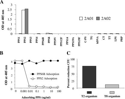
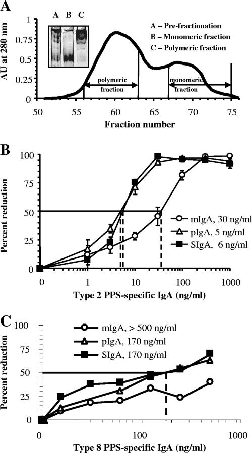
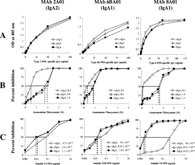
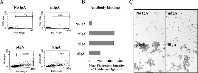
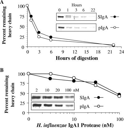
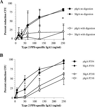
Antibodies of the immunoglobulin A (IgA) class react with capsular polysaccharides of Streptococcus pneumoniae and support complement-dependent opsonophagocytosis (OPC) of the organism by phagocytes. We characterized the biologic impact of the molecular forms of human monoclonal capsule-specific IgA (monomeric IgA [mIgA], polymeric IgA [pIgA], and secretory IgA [SIgA]) on OPC and susceptibility to cleavage by IgA1 protease. The efficiency of SIgA in support of OPC of S. pneumoniae was comparable to that of pIgA, and both forms exceeded that of mIgA by a fivefold margin. This structure-function relationship was associated with three factors. First, the avidities, or functional affinities, of both pIgA and SIgA for pneumococcal capsules exceeded those of mIgA. Second, both pIgA and SIgA required less complement to achieve similar levels of bacterial OPC than did mIgA, indicating that secretory component does not hinder the effect of complement. Third, both pIgA and SIgA mediated agglutination of the organism, whereas mIgA did not. All three forms of capsule-specific IgA showed comparable susceptibilities to cleavage and functional inhibition by bacterial IgA1 protease, demonstrating that secretory component does not prevent the proteolytic degradation of IgA1 by IgA1 protease. IgA1 cleavage results in formation of identical Fab fragments for each of the molecular forms, thereby abolishing the contribution of multivalence of pIgA and SIgA. In summary, the polymeric forms of IgA (both pIgA and SIgA) provide a substantial advantage in binding, agglutination, and OPC of the organism.


Figures

FIG. 1.
FIG. 1.
Monoclonal antibody specificities. (A) MAb 2A01 and 2A02 cross-reactivity examined using 18 coating antigens. C-PS, cell wall polysaccharide; TG, thyroglobulin; hDNA, human double-stranded DNA; CT, whole cholera toxin; TT, tetanus toxoid; LPS, lipopolysaccharide; PRP, polyribosyl ribitol phosphate. (B) Competitive inhibition ELISA using MAb 2A01 and PPS type 2 or PPS type 6B as an adsorbing antigen. (C) OPC-CFU using MAb 2A01 against two serotypes of S. pneumoniae.
FIG. 2.
FIG. 2.
(A) Affinity-purified MAb 2A01 supernatant (inset; lane A) was fractionated by gel filtration to yield mIgA (inset; lane B) and pIgA (inset; lane C). AU, absorbance units. (B) Enhanced opsonophagocytosis with MAb 2A01 (IgA2) pIgA and SIgA compared to that seen with mIgA as measured by OPC-CFU (n = 3; error bars = 1 ± standard error of the mean; key indicates the opsonophagocytic index for each molecular form). (C) MAb 8A01 showed results similar to those obtained with MAb 2A01.
FIG. 3.
FIG. 3.
Solid-phase ELISA was used to examine affinity and avidity for the three molecular forms of IgA. Antibody binding curves for mIgA, pIgA, and SIgA of one IgA2 (MAb 2A01) and two IgA1s (MAb 6BA01 and MAb 8A01). (A) ELISA binding curves show similar antigen affinities. (B) More ammonium thiocyanate is needed to disrupt pIgA and SIgA antigen binding than is required for disruption of mIgA binding for each of the three monoclonal antibodies (keys indicate the molar amounts of ammonium thiocyanate which result in a 50% reduction of antigen binding for each molecular form). (C) Lower amounts of soluble antigen are able to competitively bind 50% of pIgA and SIgA compared to the amounts of soluble antigen required for mIgA for each of the three monoclonal antibodies (keys indicate the calculated disassociation constant expressed in molar concentration for each molecular form).
FIG. 4.
FIG. 4.
Opsonophagocytic activity, binding, and phagocytosis are dependent on the presence of complement. (A) Opsonophagocytosis (OPC-CFU) was tested with 30 ng/ml 2A01 mIgA, pIgA, or SIgA at four concentrations of complement (0%, 1%, 3%, and 10%). All three forms showed a direct relationship between complement concentration and amount of opsonophagocytosis (n = 3). Asterisks designate significant differences between pIgA or SIgA and mIgA (Student's t test; P < 0.05). (B) CFU per well after OPC-CFU for a control lacking complement (light bars) or when 10% complement is present (dark bars) for each molecular form of MAb 2A01 or in the absence of antibody (n = 3; error bars = 1 ± standard error of the mean; the line across the chart at 2,000 CFU represents the starting bacterial inoculum). Control results show that the presence of complement alone did result in fewer CFU at the end of the assay, while numbers of CFU seen with a combination of complement and antibody were lower than those seen with either alone. (C) CFU per well after OPC-CFU, comparing the results obtained in the absence of complement (light bars) to those obtained with 10% heat-inactivated complement (dark bars) for each molecular form of MAb 2A01 or in the absence of antibody (n = 1; the line across the chart at 2,000 CFU represents the starting bacterial inoculum). Control findings show that heat-inactivated complement gave CFU results similar to those seen under conditions without complement, demonstrating a lack of nonspecific organism inhibitors in the complement preparation. (D) Binding and phagocytosis were tested at 30 ng/ml 2A01 mIgA, pIgA, and SIgA or in the absence of antibody at four concentrations of complement (0%, 1%, 3%, and 10%) (n = 1). OPC-flow analysis confirmed that for each molecular form the binding or phagocytosis was complement dependent. PMN, polymorphonuclear leukocyte.
FIG. 5.
FIG. 5.
SIgA and pIgA cause antibody-mediated agglutination of S. pneumoniae. (A) Bacterial agglutination measured by an increase in forward scatter (particle size) using flow cytometry was seen for hMAb 2A01 pIgA and SIgA but absent for mIgA. Percentages of bacteria which exceed single-cell values are indicated in each panel, including a control panel (No IgA). (B) Analysis of the mean fluorescent intensity of antibody binding to bacteria confirms that even though the mIgA results showed no agglutination, there was mIgA bound to the bacteria. (C) Light microscopy at ×1,000 total magnification also demonstrated the agglutinating capabilities of pIgA and SIgA.
FIG. 6.
FIG. 6.
SIgA and pIgA are similarly digested by H. influenzae type 1 IgA1 protease. (A) A 0.15-pmol volume of either hMAb 2A02 SIgA or pIgA was digested at 37°C with 0.15 pmol H. influenzae IgA1 protease for 0, 1, 3, 6, or 22 h. (B) SIgA or pIgA (10 nM) was digested at 37°C with H. influenzae type 1 IgA1 protease (2, 10, 20, or 100 nM) for 2 h. Samples were run on a 12% denaturing PAGE and silver stained, and percent remaining heavy chain was quantified by an inverse luminosity measurement in Photoshop.
FIG. 7.
FIG. 7.
IgA1 proteases from H. influenzae type 1 and S. pneumoniae inhibit IgA-mediated opsonophagocytosis of S. pneumoniae in the presence of complement. (A) Digestion of hMAb 2A02 pIgA and SIgA with H. influenzae IgA1 protease before use in opsonophagocytosis assays caused loss of the ability to effect bacterial killing compared to the results seen with undigested pIgA and SIgA. (B) Prolonged exposure of pIgA and SIgA to a protease-producing strain of S. pneumoniae (P210) inhibited IgA-mediated killing compared with the results seen with pIgA and SIgA exposed to a protease-negative mutant (P354). For each graph, data represent the results of three separate experiments. Asterisks designate significant differences between digested and nondigested antibody results (Student's t test; P < 0.05). No significant differences between the effects on capsule-specific pIgA and SIgA were seen.

References

    1. Almogren, A., B. W. Senior, L. M. Loomes, and M. A. Kerr. 2003. Structural and functional consequences of cleavage of human secretory and human serum immunoglobulin A1 by proteinases from Proteus mirabilis and Neisseria meningitidis. Infect. Immun. 71:3349-3356. - PMC - PubMed
    1. Bachmann, M. F., A. Kalinke, A. Althage, G. Freer, C. Burkhart, H.-P. Roost, H. Aguet, H. Hengartner, and R. M. Zinkernagel. 1997. The role of antibody concentration and avidity in antiviral protection. Science 276:2024-2027. - PubMed
    1. Berdoz, J., C. T. Blanc, M. Reinhardt, J. P. Kraehenbuhl, and B. Corthésy. 1999. In vitro comparison of the antigen-binding and stability properties of the various molecular forms of IgA antibodies assembled and produced in CHO cells. Proc. Natl. Acad. Sci. USA 96:3029-3034. - PMC - PubMed
    1. Berdoz, J., and B. Corthésy. 2004. Human polymeric IgA is superior to IgG and single-chain Fv of the same monoclonal specificity to inhibit urease activity associated with Helicobacter pylori. Mol. Immunol. 41:1013-1022. - PubMed
    1. Bogers, W. M. J. M., R.-K. Stad, L. A. van Es, and M. R. Daha. 1991. Immunoglobulin A: interaction with complement, phagocytic cells and endothelial cells. Complement Inflamm. 8:347-358. - PubMed

Publication types

MeSH terms