

Nucleus- and cell-specific gene expression in monkey thalamus
- PMID: 17261798
- PMCID: PMC1783903
- DOI: 10.1073/pnas.0610742104
Nucleus- and cell-specific gene expression in monkey thalamus
Abstract
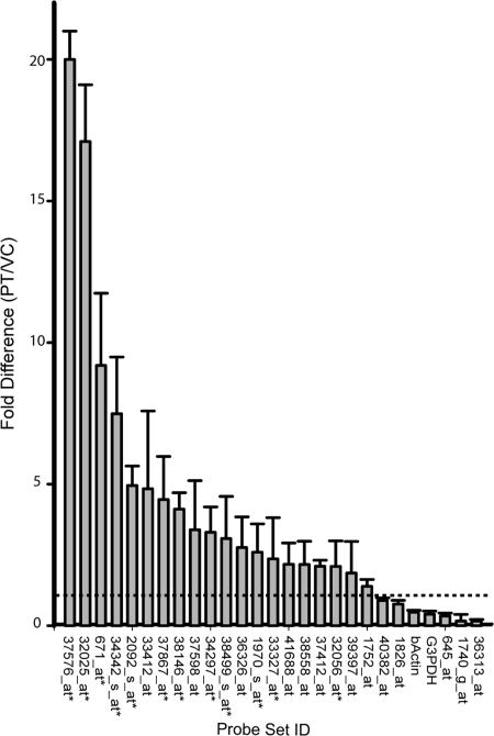
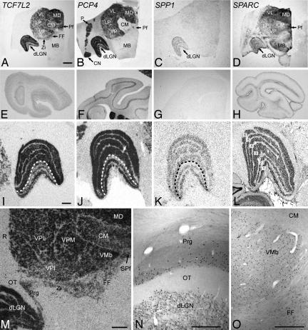
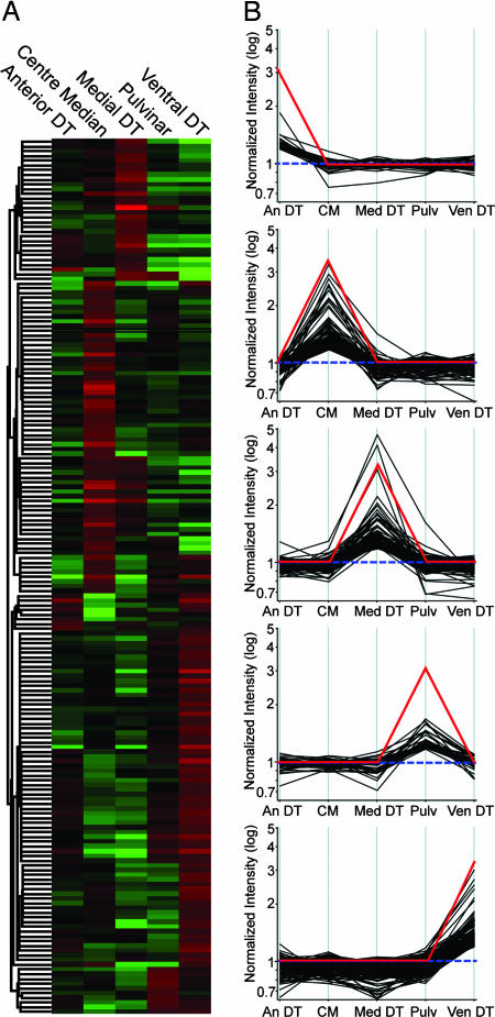
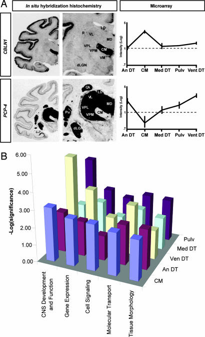
Nuclei of the mammalian thalamus are aggregations of neurons with unique architectures and input-output connections, yet the molecular determinants of their organizational specificity remain unknown. By comparing expression profiles of thalamus and cerebral cortex in adult rhesus monkeys, we identified transcripts that are unique to dorsal thalamus or to individual nuclei within it. Real-time quantitative PCR and in situ hybridization analyses confirmed the findings. Expression profiling of individual nuclei microdissected from the dorsal thalamus revealed additional subsets of nucleus-specific genes. Functional annotation using Gene Ontology (GO) vocabulary and Ingenuity Pathways Analysis revealed overrepresentation of GO categories related to development, morphogenesis, cell-cell interactions, and extracellular matrix within the thalamus- and nucleus-specific genes, many involved in the Wnt signaling pathway. Examples included the transcription factor TCF7L2, localized exclusively to excitatory neurons; a calmodulin-binding protein PCP4; the bone extracellular matrix molecules SPP1 and SPARC; and other genes involved in axon outgrowth and cell matrix interactions. Other nucleus-specific genes such as CBLN1 are involved in synaptogenesis. The genes identified likely underlie nuclear specification, cell phenotype, and connectivity during development and their maintenance in the adult thalamus.
Conflict of interest statement
The authors declare no conflict of interest.
Figures
Similar articles
-
Developmental changes in the expression of growth-associated protein-43 mRNA in the monkey thalamus: northern blot and in situ hybridization studies.Neuroscience. 2005;136(2):497-507. doi: 10.1016/j.neuroscience.2005.08.034. Epub 2005 Oct 3. Neuroscience. 2005. PMID: 16203103
-
Comparative analysis of layer-specific genes in Mammalian neocortex.Cereb Cortex. 2007 Aug;17(8):1918-33. doi: 10.1093/cercor/bhl102. Epub 2006 Oct 25. Cereb Cortex. 2007. PMID: 17065549
-
Region-specific gene expression in early postnatal mouse thalamus.J Comp Neurol. 2011 Feb 15;519(3):544-61. doi: 10.1002/cne.22532. J Comp Neurol. 2011. PMID: 21192083
-
Unveiling the diversity of thalamocortical neuron subtypes.Eur J Neurosci. 2012 May;35(10):1524-32. doi: 10.1111/j.1460-9568.2012.08033.x. Eur J Neurosci. 2012. PMID: 22606998 Review.
-
Cell nucleus in context.Crit Rev Eukaryot Gene Expr. 2000;10(1):13-20. doi: 10.1615/critreveukargeneexpr.v10.i1.30. Crit Rev Eukaryot Gene Expr. 2000. PMID: 10813390 Free PMC article. Review.
Cited by
-
Neurochemistry of the Anterior Thalamic Nuclei.Mol Neurobiol. 2017 Sep;54(7):5248-5263. doi: 10.1007/s12035-016-0077-y. Epub 2016 Aug 30. Mol Neurobiol. 2017. PMID: 27578016 Review.
-
Expression of Pcp4 gene during osteogenic differentiation of bone marrow mesenchymal stem cells in vitro.Mol Cell Biochem. 2008 Feb;309(1-2):143-50. doi: 10.1007/s11010-007-9652-x. Epub 2007 Nov 16. Mol Cell Biochem. 2008. PMID: 18008138
-
Uncovering molecular biomarkers that correlate cognitive decline with the changes of hippocampus' gene expression profiles in Alzheimer's disease.PLoS One. 2010 Apr 13;5(4):e10153. doi: 10.1371/journal.pone.0010153. PLoS One. 2010. PMID: 20405009 Free PMC article.
-
TCF7L2 polymorphisms are associated with amygdalar volume in elderly individuals with Type 2 Diabetes.Sci Rep. 2019 Nov 1;9(1):15818. doi: 10.1038/s41598-019-48899-3. Sci Rep. 2019. PMID: 31676834 Free PMC article.
-
Molecular correlates of laminar differences in the macaque dorsal lateral geniculate nucleus.J Neurosci. 2008 Nov 12;28(46):12010-22. doi: 10.1523/JNEUROSCI.3800-08.2008. J Neurosci. 2008. PMID: 19005066 Free PMC article.
References
Publication types
MeSH terms
Associated data
- Actions
Grants and funding
LinkOut - more resources
Full Text Sources
Molecular Biology Databases
Research Materials
Miscellaneous
