. 2007 Feb 6;104(6):1811-6.
doi: 10.1073/pnas.0607216104. Epub 2007 Jan 29.

The tail of the ParG DNA segregation protein remodels ParF polymers and enhances ATP hydrolysis via an arginine finger-like motif

Affiliations

The tail of the ParG DNA segregation protein remodels ParF polymers and enhances ATP hydrolysis via an arginine finger-like motif

Daniela Barillà et al. Proc Natl Acad Sci U S A. .

Abstract

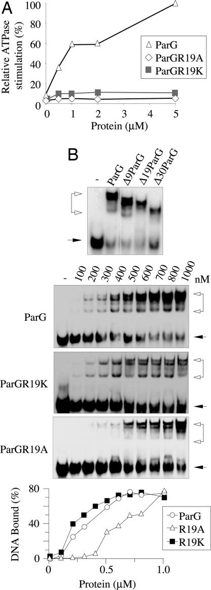
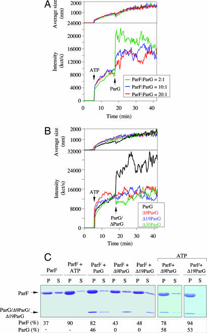
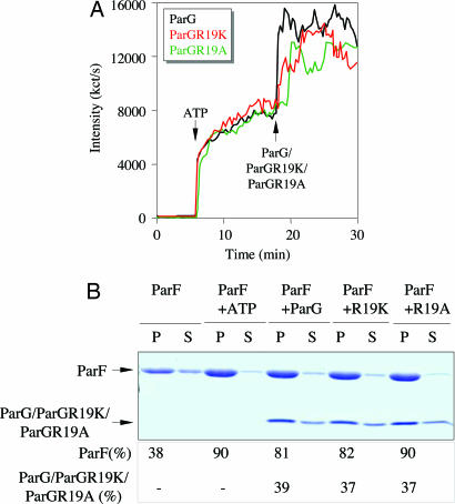
The ParF protein of plasmid TP228 belongs to the ubiquitous superfamily of ParA ATPases that drive DNA segregation in bacteria. ATP-bound ParF polymerizes into multistranded filaments. The partner protein ParG is dimeric, consisting of C-termini that interweave into a ribbon-helix-helix domain contacting the centromeric DNA and unstructured N-termini. ParG stimulates ATP hydrolysis by ParF approximately 30-fold. Here, we establish that the mobile tails of ParG are crucial for this enhancement and that arginine R19 within the tail is absolutely required for activation of ParF nucleotide hydrolysis. R19 is part of an arginine finger-like loop in ParG that is predicted to intercalate into the ParF nucleotide-binding pocket thereby promoting ATP hydrolysis. Significantly, mutations of R19 abrogated DNA segregation in vivo, proving that intracellular stimulation of ATP hydrolysis by ParG is a key regulatory process for partitioning. Furthermore, ParG bundles ParF-ATP filaments as well as promoting nucleotide-independent polymerization. The N-terminal flexible tail is required for both activities, because N-terminal DeltaParG polypeptides are defective in both functions. Strikingly, the critical arginine finger-like residue R19 is dispensable for ParG-mediated remodeling of ParF polymers, revealing that the ParG N-terminal tail possesses two separable activities in the interplay with ParF: a catalytic function during ATP hydrolysis and a mechanical role in modulation of polymerization. We speculate that activation of nucleotide hydrolysis via an arginine finger loop may be a conserved, regulatory mechanism of ParA family members and their partner proteins, including ParA-ParB and Soj-Spo0J that mediate DNA segregation and MinD-MinE that determine septum localization.


Conflict of interest statement

The authors declare no conflict of interest.

Figures

Fig. 1.
Fig. 1.
ParG N terminus is responsible for stimulation of ParF ATPase activity. (A) Domain organization of ParG and truncated proteins. Arrow, β-sheet; cylinder, α-helix; red boxes, residues R19 and R23. (B) Relative stimulation of ParF ATP hydrolysis by ParG proteins plotted as a function of their concentration. (C) E. coli two-hybrid analysis showing interaction of ParF with full-length and truncated ParG proteins. parF and parG alleles were cloned in pT18 and pT25, respectively, and analyzed as described (4). Colonies displaying an interaction are red, whereas, in the absence of interaction, they are white.
Fig. 2.
Fig. 2.
R19 in the ParG N terminus is a key residue for enhancement of ATP hydrolysis by ParF. (A) Relative stimulation of ParF ATPase activity promoted by ParG, ParGR19K, and ParGR19A plotted as a function of ParG proteins concentration. (B) Electrophoretic mobility shift assays in which a biotinylated 48-bp oligonucleotide corresponding to the parFG operator was incubated with ParG or mutant proteins. (Top) ParG and ΔParG mutants were added at 1 μM dimer. Filled arrows, free DNA; open arrows, nucleoprotein complexes.
Fig. 3.
Fig. 3.
The ParG flexible tail promotes ParF polymerization. (A) ParF polymerization followed by DLS. ParF (2.16 μM) was incubated at 30°C. ATP (500 μM) and MgCl2 (5 mM) were added at 6 min and, subsequently, ParG at ratios indicated. (Lower) Increase in light-scattering intensity expressed as kct/s. (Upper) corresponding augmentation in polymer average hydrodynamic size. (B) DLS experiment in which ParF (2.16 μM) was incubated first with ATP and MgCl2 and then with ParG or ΔParG polypeptides at a 10:1 ratio. (C) Sedimentation assay in which ParF (10 μM) was incubated without nucleotides or with ATP (2 mM) and with ParG, Δ9ParG, or Δ19ParG (10 μM). In the two rightmost panels, ParF (10 μM) was incubated with ATP (2 mM) and either Δ9ParG or Δ19ParG (10 μM). After centrifugation, 100% of pellet (P) and 33% of supernatant (S) fractions were resolved on a 15% SDS gel stained with Coomassie blue. Percentages of proteins in pellets are shown. Analogous results were obtained with Δ30ParG (data not shown).
Fig. 4.
Fig. 4.
ParGR19K and ParGR19A are as proficient as wild-type ParG in stimulating ParF filamentation. (A) DLS experiment in which ParG, ParGR19K, or ParGR19A was added to polymerizing ParF (2.16 μM) at a molar ratio of 10:1 (ParF/ParG). (B) Sedimentation assay. ParF (10 μM) was incubated without nucleotide, with ATP (2 mM) only, or with ParG, ParGR19K, or ParGR19A (10 μM). Reactions were centrifuged, and 100% of pellet (P) and 33% of supernatant (S) were resolved on a 15% SDS gel. Percentages of proteins in the pellet are shown.
Fig. 5.
Fig. 5.
Putative arginine finger residues in ParG and MinE proteins. (A) N-terminal regions of ParG homologs. Blue, putative arginine finger residue; magenta, alanine/serine patch. Region 17–23 in TP228 ParG, which is less mobile than the remainder of the N-terminal region, is boxed. Secondary structure features in TP228 ParG are shown above (6, 7). (B) N-terminal regions of MinE homologs. Conserved arginines that are candidate arginine finger residues are outlined in blue. Secondary structure features in E. coli and Neisseria meningitidis MinE are shown (21, 22, 36). The E. coli MinE N-terminal domain (residues 1–35) is predicted to consist of an extended or nascent helix (22).

References

    1. Hayes F, Barillà D. Nat Rev Microbiol. 2006;4:133–143. - PubMed
    1. Ebersbach G, Gerdes K. Annu Rev Genet. 2005;39:453–479. - PubMed
    1. Hayes F. Mol Microbiol. 2000;37:528–541. - PubMed
    1. Barillà D, Hayes F. Mol Microbiol. 2003;49:487–499. - PubMed
    1. Fothergill TJG, Barillà D, Hayes F. J Bacteriol. 2005;187:2651–2661. - PMC - PubMed

Publication types

MeSH terms

LinkOut - more resources