

Phellinus linteus activates different pathways to induce apoptosis in prostate cancer cells
- PMID: 17262078
- PMCID: PMC2360058
- DOI: 10.1038/sj.bjc.6603595
Phellinus linteus activates different pathways to induce apoptosis in prostate cancer cells
Abstract
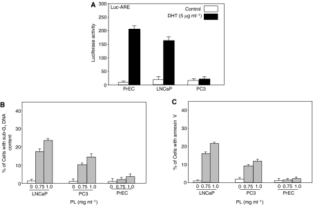
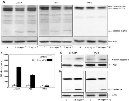
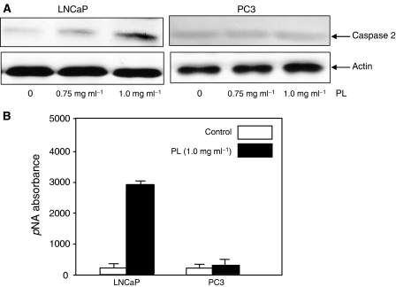
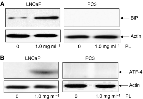
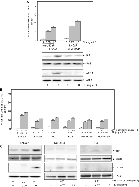
It is known that polysaccharides extracted from the Phellinus linteus (PL) mushroom possess antitumour activity. We previously have demonstrated that high doses of PL render murine or human lung cancer cells susceptible to apoptosis. However, the molecular mechanisms of PL-mediated apoptosis have not been fully explored. In this study, we demonstrate that LNCaP cells expressing the androgen receptor (AR) are highly susceptible to apoptosis in response to treatment with high doses of PL. In this process, caspase 8 and its downstream effectors (such as BID), as well as ER stress-related, apoptotic signalling, are activated. In contrast, a moderate amount of apoptosis occurs in PC3 cells (that lack AR) after the same treatment, which does not activate ER-mediated apoptotic signalling. We also show that, in the process of PL-induced apoptosis, caspase 2 is induced in LNCaP cells, but not in PC3 cells. However, LNCaP cells that express a mutated AR or LNCaP cells treated with a caspase 2 inhibitor blocked ER stress-induced apoptotic signals. The magnitudes of the induction of apoptosis in these cells are comparable with what occurred in the PC3 cells. The data demonstrate that high doses of PL activate the AR-dependent and independent apoptotic pathways. Our study also suggests that caspase 2 is a key target in the determination of the susceptibility of prostate cancer cells to PL-induced apoptosis.
Figures
Similar articles
-
Phellinus linteus sensitises apoptosis induced by doxorubicin in prostate cancer.Br J Cancer. 2006 Aug 7;95(3):282-8. doi: 10.1038/sj.bjc.6603277. Epub 2006 Jul 25. Br J Cancer. 2006. PMID: 16868541 Free PMC article.
-
Modulation of lung cancer growth arrest and apoptosis by Phellinus Linteus.Mol Carcinog. 2007 Feb;46(2):144-54. doi: 10.1002/mc.20275. Mol Carcinog. 2007. PMID: 17131292
-
Phellinus linteus extract sensitizes advanced prostate cancer cells to apoptosis in athymic nude mice.PLoS One. 2010 Mar 31;5(3):e9885. doi: 10.1371/journal.pone.0009885. PLoS One. 2010. PMID: 20360989 Free PMC article.
-
Non-genomic effects of the androgen receptor and vitamin D agonist are involved in suppressing invasive phenotype of prostate cancer cells.Steroids. 2006 Apr;71(4):304-9. doi: 10.1016/j.steroids.2005.09.010. Epub 2005 Nov 9. Steroids. 2006. PMID: 16289173 Review.
-
A medicinal mushroom: Phellinus linteus.Curr Med Chem. 2008;15(13):1330-5. doi: 10.2174/092986708784534929. Curr Med Chem. 2008. PMID: 18537612 Review.
Cited by
-
Artificial Cultivation Characteristics and Bioactive Effects of Novel Tropicoporus linteus (Syn. Phellinus linteus) Strains HN00K9 and HN6036 in Korea.Mycobiology. 2021 Apr 22;49(2):161-172. doi: 10.1080/12298093.2021.1892568. eCollection 2021. Mycobiology. 2021. PMID: 37970180 Free PMC article.
-
Optimization to the Culture Conditions for Phellinus Production with Regression Analysis and Gene-Set Based Genetic Algorithm.Biomed Res Int. 2016;2016:1358142. doi: 10.1155/2016/1358142. Epub 2016 Aug 16. Biomed Res Int. 2016. PMID: 27610365 Free PMC article.
-
Optimization to the Phellinus experimental environment based on classification forecasting method.PLoS One. 2017 Sep 28;12(9):e0185444. doi: 10.1371/journal.pone.0185444. eCollection 2017. PLoS One. 2017. PMID: 28957375 Free PMC article.
-
Mushroom-derived bioactive compounds pharmacological properties and cancer targeting: a holistic assessment.Discov Oncol. 2025 May 2;16(1):654. doi: 10.1007/s12672-025-02371-z. Discov Oncol. 2025. PMID: 40314874 Free PMC article. Review.
-
Species identity of Phellinus linteus (sanghuang) extensively used as a medicinal mushroom in Korea.J Microbiol. 2016 Apr;54(4):290-5. doi: 10.1007/s12275-016-5520-2. Epub 2016 Apr 1. J Microbiol. 2016. PMID: 27033204
References
-
- Bhornberry NA, Lazebnik Y (1998) Caspases: enemies within. Science 281: 1312–1316 - PubMed
-
- Borchers AT, Stern JS, Hackman RM, Keen CL, Gershwin EM (1999) Minireview: mushrooms, tumors and immunity. Soc Exp Biol Med 221: 281–293 - PubMed
-
- Breckenridge DG, Germain M, Mathai JP, Nguyen M, Shore GC (2003) Regulation of apoptosis by endoplasmic reticulum pathways. Oncogene 22: 8608–8618 - PubMed
-
- Chae H, Kim H, Xu C, Bailly-Maitre B, Krajewska M, Krajewski S, Banares S, Cui J, Digicaylioglu M, Ke N, Kitada S, Monosov E, Thomas M, Kress CL, Babendure JR, Tsien RY, Lipton SA, Reed JC (2004) BI-1 regulates an apoptosis pathway linked to endoplasmic reticulum stress. Mol Cell 15: 355–366 - PubMed
-
- Chen DC, Welsbie DS, Tran C, Baek SH, Chen R, Vessella R, Rosenfeld MG, Sawyers CL (2004) Molecular determinants of resistance to antiandrogen therapy. Nat Med 10: 33–39 - PubMed
Publication types
MeSH terms
Substances
Grants and funding
LinkOut - more resources
Full Text Sources
Medical
Research Materials
