

Clinically proven markers of metastasis predict metastatic spread of human melanoma cells engrafted in scid mice
- PMID: 17262079
- PMCID: PMC2360047
- DOI: 10.1038/sj.bjc.6603594
Clinically proven markers of metastasis predict metastatic spread of human melanoma cells engrafted in scid mice
Abstract
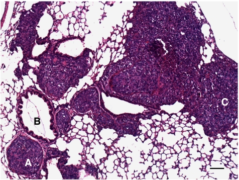
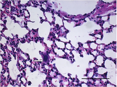
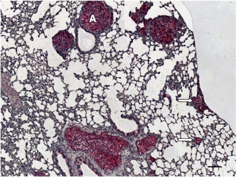
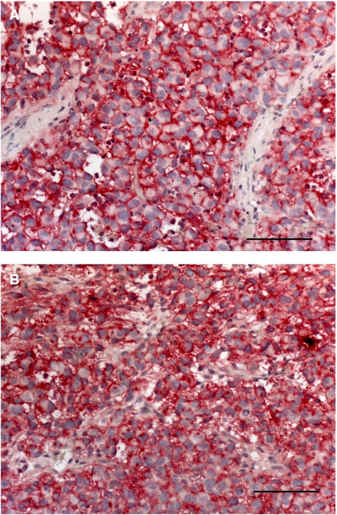
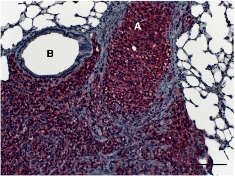
Metastasis formation is a complex process and as such can only be modelled in vivo. As markers indicating metastatic spread in syngenic mouse models differ significantly from those in man, this study aimed to develop a human melanoma xenograft mouse model that reflects the clinical situation. Six human melanoma cell lines (LOX, MV3, FEMX-1, G361, MeWo and UISO-Mel6) were xenografted into severe combined immunodeficient mice and tumour growth, metastatic behaviour and number of lung metastases were assessed. Tumours and metastases were analysed for HPA binding and expression of CEACAM-1 and L1, all markers indicative of metastasis in clinical studies. Development of primary tumour nodules ranged from 3 weeks (MV3) to 3 months (MeWo). Whereas G361 and FEMX-1 rarely formed lung metastases, MeWo, MV3 and LOX were moderately and UISO-Mel6 was highly metastatic. Similar to clinical studies, HPA, CEACAM1 and L1 indicated metastatic spread in the xenograft melanoma model, but were not all simultaneously expressed in all cell lines. Considering the strongest expression of one marker combined with an absent or low expression of the other two markers, we conclude that LOX is the cell line of choice for analyses of the functional role of HPA-binding glycoconjugates, UISO-Mel6 is ideally suited to study CEACAM1 function in melanoma spread and L1 function can best be modelled using MeWo.
Figures
Similar articles
-
Invasion potential and N-acetylgalactosamine expression in a human melanoma model.Int J Cancer. 1998 Feb 9;75(4):609-14. doi: 10.1002/(sici)1097-0215(19980209)75:4<609::aid-ijc19>3.0.co;2-3. Int J Cancer. 1998. PMID: 9466664
-
Magnetic resonance imaging of melanoma metastases in a clinical relevant human melanoma xenograft scid mouse model.Cancer Lett. 2009 Feb 18;274(2):194-200. doi: 10.1016/j.canlet.2008.09.011. Epub 2008 Oct 14. Cancer Lett. 2009. PMID: 18922631
-
A model of human cancer metastasis: extensive spontaneous and artificial metastasis of a human pigmented melanoma and derived variant sublines in nude mice.J Natl Cancer Inst. 1984 Jan;72(1):93-108. doi: 10.1093/jnci/72.1.93. J Natl Cancer Inst. 1984. PMID: 6582307
-
S100A4 expression with reduced E-cadherin expression predicts distant metastasis of human malignant melanoma cell lines in the NOD/SCID/gammaCnull (NOG) mouse model.Oncol Rep. 2005 Sep;14(3):633-7. Oncol Rep. 2005. PMID: 16077966
-
Metastases of human tumors in experimental animals.Anticancer Res. 1987 Sep-Oct;7(5B):997-1003. Anticancer Res. 1987. PMID: 3324940 Review.
Cited by
-
E- and p-selectins are essential for repopulation of chronic myelogenous and chronic eosinophilic leukemias in a scid mouse xenograft model.PLoS One. 2013 Jul 26;8(7):e70139. doi: 10.1371/journal.pone.0070139. Print 2013. PLoS One. 2013. PMID: 23922938 Free PMC article.
-
Systemic dysregulation of CEACAM1 in melanoma patients.Cancer Immunol Immunother. 2010 Feb;59(2):215-30. doi: 10.1007/s00262-009-0740-5. Cancer Immunol Immunother. 2010. PMID: 19633846 Free PMC article.
-
The Chemokine Receptor CXCR6 Evokes Reverse Signaling via the Transmembrane Chemokine CXCL16.Int J Mol Sci. 2017 Jul 8;18(7):1468. doi: 10.3390/ijms18071468. Int J Mol Sci. 2017. PMID: 28698473 Free PMC article.
-
CEACAM1 promotes melanoma metastasis and is involved in the regulation of the EMT associated gene network in melanoma cells.Sci Rep. 2018 Aug 8;8(1):11893. doi: 10.1038/s41598-018-30338-4. Sci Rep. 2018. PMID: 30089785 Free PMC article.
-
YKL-40 protein expression in human tumor samples and human tumor cell line xenografts: implications for its use in tumor models.Cell Oncol (Dordr). 2021 Oct;44(5):1183-1195. doi: 10.1007/s13402-021-00630-z. Epub 2021 Aug 25. Cell Oncol (Dordr). 2021. PMID: 34432260 Free PMC article.
References
-
- Balch CM, Buzaid AC, Atkins MB, Cascinelli N, Coit DG, Fleming ID, Houghton AJ, Kirkwood JM, Mihm MF, Morton DL, Reintgen D, Ross MI, Sober A, Soong SJ, Thompson JA, Thompson JF, Gershenwald JE, McMasters KM (2000) A new American Joint Committee on Cancer staging system for cutaneous melanoma. Cancer 88: 1484–1491 - PubMed
-
- Brooks SA, Lymboura M, Schumacher U, Leathem AJ (1996) Histochemistry to detect Helix pomatia lectin binding in breast cancer: methodology makes a difference. J Histochem Cytochem 44: 519–524 - PubMed
-
- De Giorgi V, Massi D, Germini G, Mannone F, Quercioli E, Carli P (2003) Immediate local recurrence after the excision of a polypoid melanoma: tumor dormancy or tumor activation? Dermatol Surg 29: 664–667 - PubMed
MeSH terms
Substances
LinkOut - more resources
Full Text Sources
Medical
Research Materials
Miscellaneous
