Scapular deformity in obstetric brachial plexus palsy: a new finding
- PMID: 17262175
- PMCID: PMC1820760
- DOI: 10.1007/s00276-006-0173-1
Scapular deformity in obstetric brachial plexus palsy: a new finding
Abstract
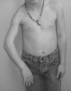
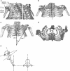
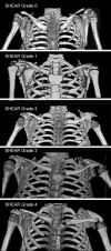
While most obstetric brachial plexus palsy patients recover arm and hand function, the residual nerve weakness leads to muscle imbalances about the shoulder which may cause bony deformities. In this paper we describe abnormalities in the developing scapula and the glenohumeral joint. We introduce a classification for the deformity which we term Scapular Hypoplasia, Elevation and Rotation. Multiple anatomic parameters were measured in bilateral CT images and three-dimensional CT reconstruction of the shoulder girdle of 30 obstetric brachial plexus palsy patients (age range 10 months-10.6 years). The affected scapulae were found to be hypoplastic by an average of 14% while the ratio of the height to the width of the body of scapula (excluding acromion) were not significantly changed, the acromion was significantly elongated by an average of 19%. These parameters as well as subluxation of the humeral head (average 14%) and downward rotation in the scapular plane were found to correlate with the area of scapula visible over the clavicle. This finding provides a classification tool for diagnosis and objective evaluation of the bony deformity and its severity in obstetric brachial plexus palsy patients.
Figures


Similar articles
-
Correlation between clinical findings and CT scan parameters for shoulder deformities in birth brachial plexus palsy.J Hand Surg Am. 2013 Aug;38(8):1557-66. doi: 10.1016/j.jhsa.2013.04.025. Epub 2013 Jun 28. J Hand Surg Am. 2013. PMID: 23816519
-
Coracoid abnormalities and their relationship with glenohumeral deformities in children with obstetric brachial plexus injury.BMC Musculoskelet Disord. 2010 Oct 13;11:237. doi: 10.1186/1471-2474-11-237. BMC Musculoskelet Disord. 2010. PMID: 20942927 Free PMC article.
-
Limited glenohumeral cross-body adduction in children with brachial plexus birth palsy: a contributor to scapular winging.J Pediatr Orthop. 2015 Apr-May;35(3):240-5. doi: 10.1097/BPO.0000000000000242. J Pediatr Orthop. 2015. PMID: 24992351
-
[Current concepts in perinatal brachial plexus palsy. Part 2: late phase. Shoulder deformities].Arch Argent Pediatr. 2011 Oct;109(5):429-36. doi: 10.5546/aap.2011.429. Arch Argent Pediatr. 2011. PMID: 22042074 Review. Spanish.
-
Perspectives on glenohumeral joint contractures and shoulder dysfunction in children with perinatal brachial plexus palsy.J Hand Ther. 2015 Apr-Jun;28(2):176-83; quiz 184. doi: 10.1016/j.jht.2014.12.001. Epub 2014 Dec 18. J Hand Ther. 2015. PMID: 25835253 Review.
Cited by
-
Scapular deformity in obstetric brachial plexus palsy and the Hueter-Volkmann law; a retrospective study.BMC Musculoskelet Disord. 2013 Mar 22;14:107. doi: 10.1186/1471-2474-14-107. BMC Musculoskelet Disord. 2013. PMID: 23522350 Free PMC article.
-
Shoulder function and anatomy in complete obstetric brachial plexus palsy: long-term improvement after triangle tilt surgery.Childs Nerv Syst. 2010 Aug;26(8):1009-19. doi: 10.1007/s00381-010-1174-2. Epub 2010 May 16. Childs Nerv Syst. 2010. PMID: 20473676 Free PMC article.
-
An MRI study on the relations between muscle atrophy, shoulder function and glenohumeral deformity in shoulders of children with obstetric brachial plexus injury.J Brachial Plex Peripher Nerve Inj. 2009 May 18;4:5. doi: 10.1186/1749-7221-4-5. J Brachial Plex Peripher Nerve Inj. 2009. PMID: 19450245 Free PMC article.
-
Arm rotated medially with supination - the ARMS variant: description of its surgical correction.BMC Musculoskelet Disord. 2009 Mar 16;10:32. doi: 10.1186/1471-2474-10-32. BMC Musculoskelet Disord. 2009. PMID: 19291305 Free PMC article.
-
Finger movement at birth in brachial plexus birth palsy.World J Orthop. 2013 Jan 18;4(1):24-8. doi: 10.5312/wjo.v4.i1.24. World J Orthop. 2013. PMID: 23362472 Free PMC article.
References
-
- Al-Qattan MM (2003) Classification of secondary shoulder deformities in obstetric brachial plexus palsy. J Hand Surg [Br] 28(5):483–486. DOI 10.1016/S0266–7681(02)00399-6 - PubMed
-
- None
- Birch R (2003) Late sequelae at the shoulder in obstetric palsy in children. In: Duparc J (eds) Shoulder. Elsevier, Paris, pp 55-200-E-210
-
- Birch R, Bonney G, Wynn Parry CB (1998) Birth lesions of the brachial plexus. In: Birch R, Bonney G, Wynn Parry CB (eds) Surgical disorders of the peripheral nerves. Shoulder, vol 3. Churchill Livingstone, New York, pp 209–233
-
- {'text': '', 'ref_index': 1, 'ids': [{'type': 'DOI', 'value': '10.1302/0301-620X.82B5.10389', 'is_inner': False, 'url': 'https://doi.org/10.1302/0301-620x.82b5.10389'}, {'type': 'PubMed', 'value': '10963171', 'is_inner': True, 'url': 'https://pubmed.ncbi.nlm.nih.gov/10963171/'}]}
- Cho TJ, Choi IH, Chung CY, Hwang JK (2000) The Sprengel deformity. Morphometric analysis using 3D-CT and its clinical relevance. J Bone Joint Surg Br 82(5):711–718 - PubMed
-
- {'text': '', 'ref_index': 1, 'ids': [{'type': 'PubMed', 'value': '1522089', 'is_inner': True, 'url': 'https://pubmed.ncbi.nlm.nih.gov/1522089/'}]}
- Friedman RJ, Hawthorne KB, Genez BM (1992) The use of computerized tomography in the measurement of glenoid version. J Bone Joint Surg Am 74(7):1032–1037 - PubMed
MeSH terms
LinkOut - more resources
Full Text Sources
Medical

