. 2007 May 1;403(3):593-601.
doi: 10.1042/BJ20070053.

The beta-cell specific transcription factor Nkx6.1 inhibits glucagon gene transcription by interfering with Pax6

Affiliations

The beta-cell specific transcription factor Nkx6.1 inhibits glucagon gene transcription by interfering with Pax6

Benoit R Gauthier et al. Biochem J. .

Abstract

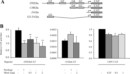
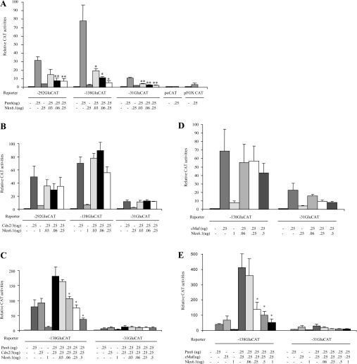
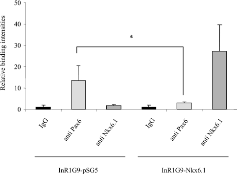
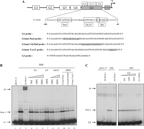
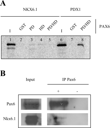
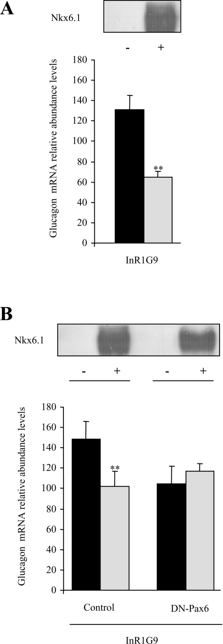
The transcription factor Nkx6.1 is required for the establishment of functional insulin-producing beta-cells in the endocrine pancreas. Overexpression of Nkx6.1 has been shown to inhibit glucagon gene expression while favouring insulin gene activation. Down-regulation resulted in the opposite effect, suggesting that absence of Nkx6.1 favours glucagon gene expression. To understand the mechanism by which Nkx6.1 suppresses glucagon gene expression, we studied its effect on the glucagon gene promoter activity in non-islet cells using transient transfections and gel-shift analyses. In glucagonoma cells transfected with an Nkx6.1-encoding vector, the glucagon promoter activity was reduced by 65%. In BHK21 cells, Nkx6.1 inhibited by 93% Pax6-mediated activation of the glucagon promoter, whereas Cdx2/3 and Maf stimulations were unaltered. Although Nkx6.1 could interact with both the G1 and G3 element, only the former displayed specificity for Nkx6.1. Mutagenesis of the three potential AT-rich motifs within the G1 revealed that only the Pax6-binding site preferentially interacted with Nkx6.1. Chromatin immunoprecipitation confirmed interaction of Nkx6.1 with the glucagon promoter and revealed a direct competition for binding between Pax6 and Nkx6.1. A weak physical interaction between Pax6 and Nkx6.1 was detected in vitro and in vivo suggesting that Nkx6.1 predominantly inhibits glucagon gene transcription through G1-binding competition. We suggest that cell-specific expression of the glucagon gene may only proceed when Nkx6.1, in combination with Pdx1 and Pax4, are silenced in early alpha-cell precursors.


Figures

Figure 1
Figure 1. Nkx6.1 inhibits trans-activation of the glucagon gene promoter in InR1G9 cells, but has no effect on a heterologous promoter
(A) Schematic representation of reporter gene constructs used in the present study. (B) Increasing amounts (0.5–2–μg) of expression vector containing the Nkx6.1 cDNA or 1–μg of expression vector containing the Pax4 cDNA were co-transfected into InR1G9 cells with 3–μg of the indicated reporter constructs. In addition, a reporter construct under the transcriptional control of the ubiquitously active CMV promoter was co-transfected into InR1G9 cells along with increasing amounts of the Nkx6.1 expression vector. Data are presented as relative basal CAT activity. **P<0.02.
Figure 2
Figure 2. Nkx6.1 interferes with Pax6-mediated transcriptional activation of the glucagon gene promoter
(A) BHK-21 cells were co-transfected with 10–μg of the indicated reporter plasmids and 0.25–μg of expression vectors encoding Pax6 either alone or with 0.03–0.25–μg of Nkx6.1 expression vector. Nkx6.1 (0.25–μg) was transfected alone as a control. (B) BHK-21 cells were co-transfected with 10–μg of the indicated reporter plasmids and 0.25–μg of expression vectors encoding Cdx2/3 either alone or with 0.03–0.25–μg of Nkx6.1 expression vector. Nkx6.1 (1–μg) was transfected alone as a control. (C) BHK-21 cells were co-transfected with 10–μg of the indicated reporter plasmids and 0.25–μg of expression vectors encoding Pax6 and 0.25–μg of Cdx2/3 either alone or with 0.03–0.5–μg of Nkx6.1 expression vector. Nkx6.1 (1–μg) was transfected alone as a control. (D) BHK-21 cells were co-transfected with 10–μg of either −138GluCAT or −31GluCAT along with 0.25–μg of expression vectors encoding c-Maf alone or in combination with 0.06–0.5–μg of Nkx6.1 expression vector. Nkx6.1 (1–μg) was transfected alone as a control. (E) BHK-21 cells were co-transfected with 10–μg of the indicated reporter plasmids and 0.25–μg of expression vectors encoding Pax6 and c-Maf either alone or with 0.06–1–μg of Nkx6.1 expression vector. Nkx6.1 (1–μg) was transfected alone as a control. Data are presented as relative CAT activity. * and ** indicate statistical significance with P<0.05 and P<0.02 respectively.
Figure 3
Figure 3. Nkx6.1 interacts with the glucagon promoter and disrupts Pax6 binding in the InR1G9 glucagon-producing cell line
Histograms representing the relative binding of Pax6 and Nkx6.1 proteins to the glucagon promoter in InR1G9 cells transfected with either an empty vector (pSG5) or the expression vector containing the Nkx6.1 cDNA. After cross-linking, immunoprecipitations were performed with anti-Pax6 or anti-Nkx6.1 as indicated in the Materials and methods section. An anti-histone H4 immunoprecipitation was also performed as a positive control in each experiment (results not shown). Binding capacity was analysed by real-time RT-PCR using a light-cycler (Roche Diagnostics). Binding intensity data are expressed in terms of the IgG immunoprecipitation (non-specific binding) and are presented as the mean±S.E.M. for at least three independent experiments.
Figure 4
Figure 4. The HD of Nkx6.1 interacts with the G1 element of the glucagon gene promoter
(A) Schematic representation of the 5′-flanking region of the rat glucagon gene promoter depicting the control elements (G1–G5). The sequence of G1 is shown as well as the binding sites for Pax6, Cdx2/3 and Maf (bold nucleotides). The various probes used in EMSA are depicted and underlined nucleotides represent mutated sequences. Studies of Nkx-6.1 binding to the G1 (−100/−49) and G3 element (−274/−234) are represented in (B) and (C) respectively. Specific interactions of Nkx6.1 on glucagon probes was determined using an anti-Nkx6.1 antibody. pBAT-12 represent nuclear extracts derived from cells transfected with control empty vector. Competition assays were performed with increasing amounts of non-labelled probes ranging from 50- to 200-fold excess for native oligonucleotides and 200-fold excess for mutated oligonucleotides. SS, supershift.
Figure 5
Figure 5. A weak interaction between Nkx6.1 and Pax6 is observed both in vitro and in vivo
(A) GST-precipitation assay using 10–μg of GST alone, GST-Pax6 PD, HD or PDHD (paired-linker-HD) fusion proteins immobilized on sepharose beads and in vitro synthesized, 35S-labelled Nkx6.1 or Pdx1. Lane input, 10% of the respective in vitro translation reaction used for protein–protein interactions. (B) To confirm in vitro interactions, InR1G9 cells were transfected with Nkx6.1 and co-immunoprecipitation was performed using an anti-mouse Pax6 serum in the presence of sheep anti-mouse IgG magnetic Dynabeads (+). Experiments were also performed in the absence of the Pax6 serum (−). Precipitates were subjected to Western blotting for Pax6 and Nkx6.1. The input for Pax6 was 50–μg while 10–μg was used for Nkx6.1.
Figure 6
Figure 6. Overexpression of Nkx6.1 inhibits endogenous levels of glucagon mRNA in InR1G9 cells by competing with Pax6 trans-activation
(A) InR1G9 cells were transiently transfected with an expression vector encoding Nkx6.1 using the TransFectin™ reagent. This approach permitted 75% transfection efficiency as assessed by FACS analysis. Expression of the Nkx6.1 transgene in transfected cells (+) and its absence in untransfected cells (−) was determined by semi-quantitative PCR. Relative abundance levels of endogenous glucagon mRNA were then evaluated by quantitative PCR and normalized to the housekeeping transcript TBP. (B) Two stable clonal derivatives of the InR1G9 cell line expressing either an empty expression vector (control) or a dominant-negative variant of Pax6 (DN-Pax6) were transiently transfected with Nkx6.1. Expression of Nkx6.1 in non-transfected (−) and transfected (+) cells was evaluated by semi-quantitative PCR. Relative abundance levels of endogenous glucagon mRNA were then evaluated by quantitative PCR and normalized to the housekeeping transcript TBP. ** indicates statistical significance with P<0.02.

References

    1. Habener J. F., Kemp D. M., Thomas M. K. Minireview: transcriptional regulation in pancreatic development. Endocrinology. 2005;146:1025–1034. - PubMed
    1. Johnson J., Carisson L., Edlund T., Edlund H. Insulin-promoter-factor 1 is required for pancreas development in mice. Nature. 1994;371:606–609. - PubMed
    1. Li H., Arber S., Jessel T. M., Edlund H. Selective agenesis of the dorsal pancreas in mice lacking homeobox gene Hlxb9. Nat. Genet. 1999;23:67–70. - PubMed
    1. Harrison K. A., Thaler J., Pfaff S. L., Gu H., Kehrl J. H. Pancreas dorsal lobe agenesis and abnormal islets of Langerhans in Hlxb9-deficient mice. Nat. Genet. 1999;23:71–75. - PubMed
    1. Gradwohl G., Dierich A., LeMeur M., Guillemot F. Neurogenin3 is required for the development of the four endocrine cell lineages of the pancreas. Proc. Natl. Acad. Sci. U.S.A. 2000;97:1607–1611. - PMC - PubMed

Publication types

MeSH terms