Activation of the galanin receptor 2 (GalR2) protects the hippocampus from neuronal damage
- PMID: 17263796
- PMCID: PMC2705497
- DOI: 10.1111/j.1471-4159.2006.04239.x
Activation of the galanin receptor 2 (GalR2) protects the hippocampus from neuronal damage
Abstract
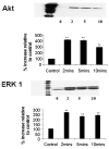
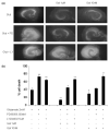
Expression of the neuropeptide galanin is up-regulated in many brain regions following nerve injury and in the basal forebrain of patients with Alzheimer's disease. We have previously demonstrated that galanin modulates hippocampal neuronal survival, although it was unclear which receptor subtype(s) mediates this effect. Here we report that the protective role played by galanin in hippocampal cultures is abolished in animals carrying a loss-of-function mutation in the second galanin receptor subtype (GalR2-MUT). Exogenous galanin stimulates the phosphorylation of the serine/threonine kinase Akt and extracellular signal-regulated kinase (ERK) in wild-type (WT) cultures by 435 +/- 5% and 278 +/- 2%, respectively. The glutamate-induced activation of Akt was abolished in cultures from galanin knockout animals, and was markedly attenuated in GalR2-MUT animals, compared with WT controls. In contrast, similar levels of glutamate-induced ERK activation were observed in both loss-of-function mutants, but were further increased in galanin over-expressing animals. Using specific inhibitors of either ERK or Akt confirms that a GalR2-dependent modulation in the activation of the Akt and ERK signalling pathways contributes to the protective effects of galanin. These findings imply that the rise in endogenous galanin observed either after brain injury or in various disease states is an adaptive response that reduces apoptosis by the activation of GalR2, and hence Akt and ERK.
Figures


Similar articles
-
Mice deficient for galanin receptor 2 have decreased neurite outgrowth from adult sensory neurons and impaired pain-like behaviour.J Neurochem. 2006 Nov;99(3):1000-10. doi: 10.1111/j.1471-4159.2006.04143.x. J Neurochem. 2006. PMID: 17076662 Free PMC article.
-
Endogenous galanin protects mouse hippocampal neurons against amyloid toxicity in vitro via activation of galanin receptor-2.J Alzheimers Dis. 2011;25(3):455-62. doi: 10.3233/JAD-2011-110011. J Alzheimers Dis. 2011. PMID: 21471641 Free PMC article.
-
Rap1 mediates galanin receptor 2-induced proliferation and survival in squamous cell carcinoma.Cell Signal. 2011 Jul;23(7):1110-8. doi: 10.1016/j.cellsig.2011.02.002. Epub 2011 Feb 21. Cell Signal. 2011. PMID: 21345369 Free PMC article.
-
Palmitate differentially regulates Spexin, and its receptors Galr2 and Galr3, in GnRH neurons through mechanisms involving PKC, MAPKs, and TLR4.Mol Cell Endocrinol. 2020 Dec 1;518:110991. doi: 10.1016/j.mce.2020.110991. Epub 2020 Aug 22. Mol Cell Endocrinol. 2020. PMID: 32841709 Review.
-
Regulation of limbic status epilepticus by hippocampal galanin type 1 and type 2 receptors.Neuropeptides. 2005 Jun;39(3):277-80. doi: 10.1016/j.npep.2004.12.003. Epub 2005 Jan 28. Neuropeptides. 2005. PMID: 15944022 Review.
Cited by
-
Conformational signatures in β-arrestin2 reveal natural biased agonism at a G-protein-coupled receptor.Commun Biol. 2018 Sep 3;1:128. doi: 10.1038/s42003-018-0134-3. eCollection 2018. Commun Biol. 2018. PMID: 30272007 Free PMC article.
-
Regulation of neurological and neuropsychiatric phenotypes by locus coeruleus-derived galanin.Brain Res. 2016 Jun 15;1641(Pt B):320-37. doi: 10.1016/j.brainres.2015.11.025. Epub 2015 Nov 20. Brain Res. 2016. PMID: 26607256 Free PMC article. Review.
-
Galanin Regulates Myocardial Mitochondrial ROS Homeostasis and Hypertrophic Remodeling Through GalR2.Front Pharmacol. 2022 Mar 31;13:869179. doi: 10.3389/fphar.2022.869179. eCollection 2022. Front Pharmacol. 2022. PMID: 35431947 Free PMC article.
-
The role of neuropeptides in suicidal behavior: a systematic review.Biomed Res Int. 2013;2013:687575. doi: 10.1155/2013/687575. Epub 2013 Aug 6. Biomed Res Int. 2013. PMID: 23986909 Free PMC article.
-
Inflammation-induced changes in the chemical coding pattern of colon-projecting neurons in the inferior mesenteric ganglia of the pig.J Mol Neurosci. 2012 Feb;46(2):450-8. doi: 10.1007/s12031-011-9613-4. Epub 2011 Aug 9. J Mol Neurosci. 2012. PMID: 21826392
References
-
- Alessi DR, Cuenda A, Cohen P, Dudley DT, Saltiel AR. PD 098059 is a specific inhibitor of the activation of mitogen-activated protein kinase kinase in vitro and in vivo. J. Biol. Chem. 1995;270:27 489–27 494. - PubMed
-
- Almeida RD, Manadas BJ, Melo CV, Gomes JR, Mendes CS, Graos MM, Carvalho RF, Carvalho AP, Duarte CB. Neuroprotection by BDNF against glutamate-induced apoptotic cell death is mediated by ERK and PI3-kinase pathways. Cell Death Differentiation. 2005;12:1329–1343. - PubMed
-
- Bacon A, Holmes FE, Small CJ, Ghatei M, Mahoney S, Bloom SR, Wynick D. Transgenic over-expression of galanin in injured primary sensory neurons. Neuroreport. 2002;13:2129–2132. - PubMed
-
- Brecht S, Buschmann T, Grimm S, Zimmermann M, Herdegen T. Persisting expression of galanin in axotomized mamillary and septal neurons of adult rats labeled for c-Jun and NADPH-diaphorase. Brain Res. Mol. Brain Res. 1997;48:7–16. - PubMed
-
- Burgevin MC, Loquet I, Quarteronet D, Habert-Ortoli E. Cloning, pharmacological characterization, and anatomical distribution of a rat cDNA encoding for a galanin receptor. J. Mol. Neurosci. 1995;6:33–41. - PubMed
Publication types
MeSH terms
Substances
Grants and funding
LinkOut - more resources
Full Text Sources
Other Literature Sources
Molecular Biology Databases
Miscellaneous

