. 2007 Jan 30:7:9.
doi: 10.1186/1471-2180-7-9.

Dengue virus type 2: replication and tropisms in orally infected Aedes aegypti mosquitoes

Affiliations

Dengue virus type 2: replication and tropisms in orally infected Aedes aegypti mosquitoes

Ma Isabel Salazar et al. BMC Microbiol. .

Abstract

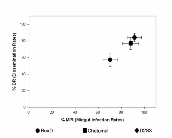
Background: To be transmitted by its mosquito vector, dengue virus (DENV) must infect midgut epithelial cells, replicate and disseminate into the hemocoel, and finally infect the salivary glands, which is essential for transmission. The extrinsic incubation period (EIP) is very relevant epidemiologically and is the time required from the ingestion of virus until it can be transmitted to the next vertebrate host. The EIP is conditioned by the kinetics and tropisms of virus replication in its vector. Here we document the virogenesis of DENV-2 in newly-colonized Aedes aegypti mosquitoes from Chetumal, Mexico in order to understand better the effect of vector-virus interactions on dengue transmission.

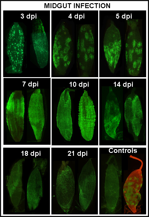
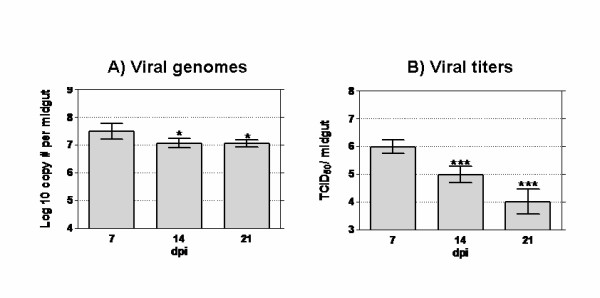
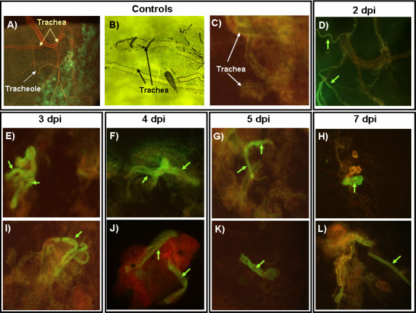
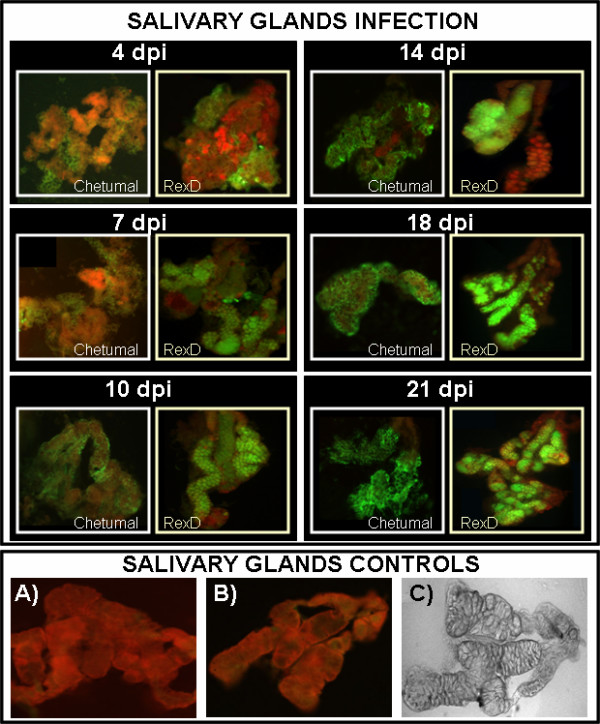
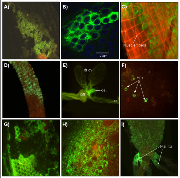
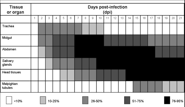
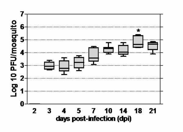
Results: After ingestion of DENV-2, midgut infections in Chetumal mosquitoes were characterized by a peak in virus titers between 7 and 10 days post-infection (dpi). The amount of viral antigen and viral titers in the midgut then declined, but viral RNA levels remained stable. The presence of DENV-2 antigen in the trachea was positively correlated with virus dissemination from the midgut. DENV-2 antigen was found in salivary gland tissue in more than a third of mosquitoes at 4 dpi. Unlike in the midgut, the amount of viral antigen (as well as the percent of infected salivary glands) increased with time. DENV-2 antigen also accumulated and increased in neural tissue throughout the EIP. DENV-2 antigen was detected in multiple tissues of the vector, but unlike some other arboviruses, was not detected in muscle.

Conclusion: Our results suggest that the EIP of DENV-2 in its vector may be shorter that the previously reported and that the tracheal system may facilitate DENV-2 dissemination from the midgut. Mosquito organs (e.g. midgut, neural tissue, and salivary glands) differed in their response to DENV-2 infection.


Figures

Figure 1
Figure 1
DENV-2 midgut infection (MIR) and dissemination rates (DR) in Aedes aegypti mosquitoes. MIRs and DRs for Chetumal, Rex-D, and D2S3 mosquito strains at 14 dpi as described in Materials and Methods. The mean and standard deviation values were obtained from four independent experiments (n= 50–60 mosquitoes).
Figure 2
Figure 2
Kinetics of DENV-2 midgut infection after oral challenge of Aedes aegypti. Chetumal mosquito midguts dissected at different time points (n = 30 for each time point) were assayed by IFA to detect DENV-2 viral antigen (green). At each time point two representative midguts from Chetumal mosquitoes are presented. Controls are an uninfected midgut and a dengue infected midgut (in red midgut musculature). The magnification was 100×.
Figure 3
Figure 3
Quantification of DENV-2 RNA and infectious virus in midguts revealed significant differences. Midguts (n = 15) from orally infected Chetumal mosquitoes were assayed to quantify viral RNA by Q-PCR and infectious particles by end point titrations at different time points. A) Viral RNA decreased slightly between 7 and 14 dpi and then remained stable, * P < 0.05. B) Viral titers declined significantly at 14 and 21 dpi, *** P < 0.0001.
Figure 4
Figure 4
DENV-2 antigen in the tracheal system of Chetumal mosquitoes following infection. DENV-2 antigen was detected by IFA in trachea of Chetumal mosquitoes at 2, 3, 4, 5, and 7 dpi (n= 40–50). Midgut- associated tracheal system (A) and uninfected trachea using light (B) and fluorescence (C) microscopy. Green arrows point to portions of DENV-2 infected trachea (E-L) (200×).
Figure 5
Figure 5
DENV-2 tropism for Aedes aegypti salivary glands. Salivary glands were dissected at the time points noted, fixed, and assayed for virus antigen by IFA (n = 30–50). Representative salivary glands (200×) from Chetumal and Rex-D mosquitoes at each time point are shown. DENV-2 virus antigen increased in the salivary glands through out the EIP. Negative controls (A and B) and salivary glands examined with light microscopy (C) are shown. In Rex-D mosquitoes, at 4 dpi viral antigen was typically associated with the fat body surrounding the salivary glands. In contrast salivary gland tissues of Chetumal mosquitoes contained viral antigen at 4 dpi.
Figure 6
Figure 6
DENV-2 tropisms in different tissues and organs of Aedes aegypti. DENV-2 viral E antigen distribution in different tissues and organs was revealed by IFA (green for FITC or Alexa 488) in Chetumal mosquitoes (n = 20). A) DENV-2 antigen in fat body of mosquito abdomen at 2 dpi. B) DENV-2 infected epithelial cells in midgut (4 dpi). C) DENV-2 is present in epithelial cells but absent in midgut-associated muscle (red phalloidin-Alexa® 546). D) DENV-2 in anterior midgut at 5 dpi. E) Infected esophagus at 7 dpi, dr.dv. dorsal diverticulum, car: cardia, oe: esophagus, and cr: crop. F) Hemocytes infected with DENV-2 at 10 dpi. G) Ommatidia of the compound eye exhibiting viral antigen at 12 dpi. H) Nervous system tissue profusely infected by DENV-2 at 14 dpi. I) Malphigian tubules expressing viral antigen (Mal. Tu.) at 16 dpi, il: ileum. These pictures represent the infection patterns observed. Original magnification was 200×, but pictures were cropped in Adobe Photoshop to improve presentation.
Figure 7
Figure 7
Temporal tropisms of DENV-2 in Chetumal mosquitoes. Infection rates in the respective organ or tissue are shown by the gray scale. Percentages were obtained after dissecting, staining and analyzing individual mosquitoes (n = 20–30) at each time point. Mosquitoes were assayed at 1, 2, 3, 5, 7, 10, 14, 18 and 21 dpi.
Figure 8
Figure 8
Kinetics of DENV-2 replication in orally infected mosquitoes. DENV-2 titers in individual Chetumal mosquitoes were determined by plaque assay at 3, 4, 7, 10, 14, 18 and 21 dpi. Each box in the graph represents mean and range values of viral titers (n = 10) in mosquitoes for each time point.

Similar articles

Cited by

References

    1. Derouich M, Boutayeb A, Twizell EH. A model of dengue fever. Biomed Eng Online. 2003;2:4. doi: 10.1186/1475-925X-2-4. - DOI - PMC - PubMed
    1. Gubler DJ. Dengue and dengue hemorrhagic fever. Clin Microbiol Rev. 1998;11:480–496. - PMC - PubMed
    1. Gubler DJ, Clark AG. Dengue/dengue hemorrhagic fever: The emergence of a global health problem. Emerg Infect Dis. 1995;1:55–57. - PMC - PubMed
    1. Holmes EC, Twiddy SS. The origin, emergence and evolutionary genetics of dengue virus. Infect Genet Evol. 2003;3:19–28. doi: 10.1016/S1567-1348(03)00004-2. - DOI - PubMed
    1. Watts DM, Burke DS, Harrison BA, Whitmire RE, Nisalak A. Effect of temperature on the vector efficiency of Aedes aegypti for dengue 2 virus. Am J Trop Med Hyg. 1987;36:143–152. - PubMed

Publication types

LinkOut - more resources