. 2007;35(4):1145-54.
doi: 10.1093/nar/gkm001. Epub 2007 Jan 30.

DNA binding mechanism revealed by high resolution crystal structure of Arabidopsis thaliana WRKY1 protein

Affiliations

DNA binding mechanism revealed by high resolution crystal structure of Arabidopsis thaliana WRKY1 protein

Ming-Rui Duan et al. Nucleic Acids Res. 2007.

Abstract

WRKY proteins, defined by the conserved WRKYGQK sequence, are comprised of a large superfamily of transcription factors identified specifically from the plant kingdom. This superfamily plays important roles in plant disease resistance, abiotic stress, senescence as well as in some developmental processes. In this study, the Arabidopsis WRKY1 was shown to be involved in the salicylic acid signaling pathway and partially dependent on NPR1; a C-terminal domain of WRKY1, AtWRKY1-C, was constructed for structural studies. Previous investigations showed that DNA binding of the WRKY proteins was localized at the WRKY domains and these domains may define novel zinc-binding motifs. The crystal structure of the AtWRKY1-C determined at 1.6 A resolution has revealed that this domain is composed of a globular structure with five beta strands, forming an antiparallel beta-sheet. A novel zinc-binding site is situated at one end of the beta-sheet, between strands beta4 and beta5. Based on this high-resolution crystal structure and site-directed mutagenesis, we have defined and confirmed that the DNA-binding residues of AtWRKY1-C are located at beta2 and beta3 strands. These results provided us with structural information to understand the mechanism of transcriptional control and signal transduction events of the WRKY proteins.


Figures

Figure 1.
Figure 1.
AtWRKY1 is partially NPR1-dependent in SA pathway. Four weeks old wild type (Col-0), npr1-3 and 35S::NPR1 Arabidopsis were sprayed with 2 mM SA and harvested at indicated time points. PR1 transcripts were detected the same as AtWRKY1 except for the difference of probe. The ethidium bromide stain of rRNA is shown for each lane to allow assessment of equal loading.
Figure 2.
Figure 2.
Structure of AtWRKY1-C and multi-sequence alignment. (A) Ribbon representation of AtWRKY1-C domain. The AtWRKY1-C is composed of five β-strands (yellow ribbons), which are numbered from the N-terminus. The zinc ion is shown as a purple sphere and the zinc-coordinating residues are represented by sticks (yellow for C, red for O, blue for N, orange for S). (B) 3D-superimposition of the structure of AtWRKY1-C and the best representative NMR structure of AtWRKY4-C (model 15), using LSQ Fit in O. Macromolecular structures are shown by cartoons, with the AtWRKY1-C colored in yellow and AtWRKY4-C in cyan. The zinc ions are represented as spheres shown by magenta in AtWRKY1-C and orange in AtWRKY4-C. (C) Charge distribution on AtWRKY1-C structure surfaces given by GRASP. Positive charges are represented by blue, negative charges are represented by red. (D) Structure-based sequence alignment of both N-terminus and C-terminus of the nine Arabidopsis WRKY proteins from group I. The zinc-coordinating residues are shown on blue background. Conserved residue elements for stabilizing the structure and recognizing DNA are drawn on red and yellow background, respectively. Residues of β1, highlighted in green but missed in AtWRKY4-C structure, are rather conserved in all C-terminal domains of group I WRKY proteins. Residues marked by stars and triangles are scaffolds of two stable regions. Details of the interactions of the two regions are represented in Figure 3C and D.
Figure 3.
Figure 3.
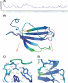
B-factor representation of AtWRKY1-C crystal structure. All the objects are colored according to the B-factor values, from blue to red in the order of increasing B-factor. (A) Plot of B-factor against residues, with B-factor averaged over each residue. (B) The overall structure of AtWRKY1-C in ribbon colored according to the B-factor values. The ends of the loop between strands β1 and β2 with low B-factor values are circled out by red rings for further amplification in 3C and 3D. Details of the interaction around these two terminals are described in 3C and 3D. (C) A D308-W312-K341 triad in the C-terminal of the loop between β1 and β2. Asp308 forms a well-defined salt-bridge with Lys341, extensively H-bonding with side chains of Trp312, Tyr357 and backbone of Tyr310. The three key residues Asp308, Trp312 and Lys341, components of D308-W312-K341 triad, and their hydrogen bonded residues Gly309, Tyr357 are marked by stars in Figure 2D. (D) The stable N-terminal end of the loop between β1 and β2. Arg345 is hydrogen bonded with the backbones of Thr301, Phe303 and Thr350. A potential salt-bridge between Arg345 and Asp304 was also observed as shown, and these two conserved residues are marked by triangles in Figure 2D.
Figure 4.
Figure 4.
The zinc coordination environment. (A) 3Fo-2Fc electron-density map. The coordination of the zinc ion by Cys332, Cys337, His361 and His363 was shown. The map was prepared using CCP4 program suite and displayed with Pymol at the contour of 1.5σ level. The zinc ion is shown as a purple sphere and the zinc-coordinating residues are represented by stick (yellow for C, red for O, blue for N, orange for S, respectively). (B) The distances between the zinc ion and the coordinate residues. The orientation and colors are the same as shown in Figure 4A.
Figure 5.
Figure 5.
EMSA with the consensus W-box binding site and wild-type AtWRKY1-C as well as mutant proteins. (A) Binding activity examination with mutations on β2. The binding reaction contained 5 μg purified proteins and 3 μg DNA duplex. The gel was stained with EB after the electrophoresis. The same amount of purified proteins were run on SDS–PAGE and stained with Coomassie Brilliant Blue to show the equal loading. (B) Binding activity examination with mutations on β3 and β4.
Figure 6.
Figure 6.
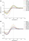
CD spectra. CD spectra of wild-type AtWRKY1-C and the mutant proteins on β2 strand. The different proteins were distinguished from each other by colors. (A) CD spectra of wild-type AtWRKY1-C and the mutant proteins on β3 and β4 strands.
Figure 7.
Figure 7.
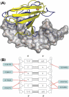
A model of the AtWRKY1-C and W-box interaction. (A) Superimposition of AtWRKY1-C and the overlapping structure of the Drosophila GCM domain using LSQ Fit in the program O. Macromolecular structures are shown by cartoon loops, with the GCM domain colored yellow, AtWRKY1-C blue and zinc ions as orange and magenta spheres. The DNA double-helix is shown with sticks wrapped inside surface rendering. (B) The final schematic model of the specific interaction of AtWRKY1-C with the W-box, combining all information obtained from the structural and mutagenesis experiments presented in this work.

References

    1. Ulker B, Somssich IE. WRKY transcription factors: from DNA binding towards biological function. Curr. Opin. Plant Biol. 2004;7:491–498. - PubMed
    1. Zhang Y, Wang L. The WRKY transcription factor superfamily: its origin in eukaryotes and expansion in plants. BMC Evol. Biol. 2005;5:1. - PMC - PubMed
    1. Chen C, Chen Z. Potentiation of developmentally regulated plant defense response by AtWRKY18, a pathogen-induced Arabidopsis transcription factor. Plant Physiol. 2002;129:706–716. - PMC - PubMed
    1. Robatzek S, Somssich IE. Targets of AtWRKY6 regulation during plant senescence and pathogen defense. Genes Dev. 2002;16:1139–1149. - PMC - PubMed
    1. Yoda H, Ogawa M, Yamaguchi Y, Koizumi N, Kusano T, Sano H. Identification of early-responsive genes associated with the hypersensitive response to tobacco mosaic virus and characterization of a WRKY-type transcription factor in tobacco plants. Mol. Genet. Genomics. 2002;267:154–161. - PubMed

Publication types

MeSH terms

Associated data