. 2007 May 15;109(10):4415-23.
doi: 10.1182/blood-2006-09-047076. Epub 2007 Jan 30.

Statins synergistically potentiate 7-hydroxystaurosporine (UCN-01) lethality in human leukemia and myeloma cells by disrupting Ras farnesylation and activation

Affiliations

Statins synergistically potentiate 7-hydroxystaurosporine (UCN-01) lethality in human leukemia and myeloma cells by disrupting Ras farnesylation and activation

Yun Dai et al. Blood. .

Abstract

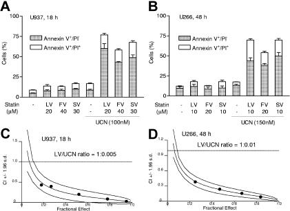
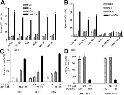
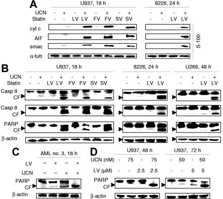
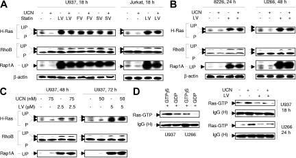
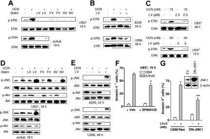
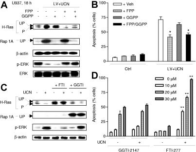
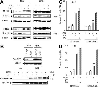
Interactions between UCN-01 and HMG-CoA reductase inhibitors (ie, statins) have been examined in human leukemia and myeloma cells. Exposure of U937 and U266 cells to minimally toxic concentrations of UCN-01 and various statins (eg, lovastatin, simvastatin, or fluvastatin) dramatically increased mitochondrial dysfunction, caspase activation, and apoptosis. Comparable effects were observed in other leukemia and myeloma cell lines as well as in primary acute myeloid leukemia (AML) blasts but not in normal hematopoietic cells. Potentiation of UCN-01 lethality by lovastatin was associated with disruption of Ras prenylation and activation. These events were significantly attenuated by farnesyl pyrophosphate (FPP) but not by geranylgeranyl pyrophosphate (GGPP), implicating perturbations in farnesylation rather than geranylgeranylation in synergistic interactions. Coexposure to statins and UCN-01 resulted in inactivation of ERK1/2 and Akt, accompanied by JNK activation. U266 cells ectopically expressing JNK1-APF, a dominant negative JNK1 mutant, displayed significantly reduced susceptibility to lovastatin/UCN-01-mediated lethality. Moreover, transfection of U266 cells with constitutively activated H-Ras (Q61L) attenuated ERK1/2 inactivation and dramatically diminished the lethality of this regimen. Collectively, these findings indicate that HMG-CoA reductase inhibitors act through a Ras farnesylation-associated mechanism to induce signaling perturbations, particularly prevention of Ras and ERK1/2 activation, in UCN-01-treated cells, resulting in the synergistic induction of cell death.


Figures

Figure 1
Figure 1
Statins interact synergistically with UCN-01 to induce apoptosis in human malignant hematopoietic cells. (A) Human myelomonoyctic leukemia U937 cells were exposed for 18 hours to 100 nM UCN-01 (UCN) with or without 20 μM lovastatin (LV), 40 μM fluvastatin (FV), or 30 μM simvastatin (SV), respectively, after which the percentage of annexin V+ cells exhibiting either annexin V+/PI (early apoptosis) or annexin V+/PI+ (late apoptosis) was determined by annexin V–FITC/PI staining and flow cytometry as described in “Analysis of apoptosis and clonogenicity.” (B) Human myeloma U266 cells were exposed for 48 hours to 150 nM UCN-01 with or without 10 μM lovastatin, 20 μM fluvastatin (FV), or 10 μM simvastatin (SV), respectively, after which the percentage of apoptotic cells was determined as described in panel A. For panels A and B, results represent the means ± SD for 3 separate experiments performed in triplicate. (C-D) U937 and U266 cells were exposed to a range of lovastatin (5 to 25 μM) and UCN-01 (50 to 250 nM) concentrations alone and in combination at a fixed ratio (eg, U937, 1:0.005; U266, 1:0.01) for 18 hours (U937) or 48 hours (U266). At the end of the exposure intervals, the percentage of annexin V+ cells was determined for each condition; fractional effect values were determined by comparing results with those of untreated controls, and median dose effect analysis was employed to characterize the nature of the interaction between lovastatin and UCN-01. CI values less than 1.0 (horizontal line) denote a synergistic interaction. The results of representative experiments are shown; 2 additional studies yielded equivalent results.
Figure 2
Figure 2
The statin/UCN-01 regimen induces apoptosis in multiple leukemia and myeloma cell lines as well as primary AML cells but is minimally toxic to normal hematopoietic cells. (A) Human leukemia (Jurkat, HL-60, and Raji) and myeloma (RPMI8226 [8226], MM.1S, and MM.1R) cells were exposed to UCN-01 ([UCN] 100 nM for HL-60, RPMI8226, MM.1S, and MM.1R; 150 nM for Jurkat and Raji) with or without lovastatin ([LV] 20 μM for all leukemia cell lines; 10 μM for all myeloma cell lines) for 18 hours (leukemia cells) or 24 hours (myeloma cells), after which the percentage of annexin V+ cells was determined by flow cytometry. (B) AML blasts were isolated from peripheral blood samples derived from 3 patients with AML (FAB classification M2) as described in “Cells and reagents.” Mononuclear cells were isolated from the bone marrow (BM/MC) of 2 patients with nonmalignant, nonmyeloid hematopoietic disorders and the peripheral blood (PB/MC) from 1 healthy volunteer. AML blasts and normal cells were treated with 150 nM UCN-01 with or without 20 μM lovastatin for 18 hours, after which the percentage of apoptotic cells was determined by evaluating Giemsa-Wright–stained cytospin preparations. (C) U937 cells were incubated with UCN-01 (50 to 100 nM) with or without low doses of lovastatin (1 to 5 μM) for 48 hours or 72 hours, after which the percentage of annexin V+ cells, including annexin V+/PI and annexin V+/PI+, was determined by flow cytometry. (D) U937 and U266 cells were treated with UCN-01 (U937, 100 nM; U266, 150 nM) with or without lovastatin (U937, 20 μM; U266, 10 μM) for 18 hours (U937) or 48 hours (U266), after which cells were washed free of drug and plated in soft agar as described in “Analysis of apoptosis and clonogenicity.” After incubation for 12 days, colonies (more than 50 cells) were scored, and colony formation for each condition expressed relative to untreated control cells. For all panels, results represent the means ± SD for 3 separate experiments performed in triplicate.
Figure 3
Figure 3
Statins potentiate mitochondrial dysfunction and activation of caspase cascades mediated by UCN-01 in cultured leukemia and myeloma cells as well as in primary AML blasts. (A) U937 cells were exposed for 18 hours to 100 nM UCN-01 (UCN) with or without 20 μM lovastatin (LV), 40 μM fluvastatin (FV), or 30 μM simvastatin (SV) (left panels), and RPMI8226 cells (8226) for 24 hours to 100 nM UCN-01 with or without 10 μM lovastatin (right panels). At the end of the incubation period, expression of cytochrome c, Smac/DIABLO, and AIF in cytosolic fractions (S-100) was evaluated by Western blot analysis as described in “Western blot analysis.” (B) U937 (left panels) and RPMI8226 cells (middle panels) were treated as described in panel A. U266 cells were exposed for 48 hours to 150 nM UCN-01 with or without 10 μM lovastatin (right panels). Cells were then lysed and subjected to Western blot analysis to assess cleavage of caspases and degradation of PARP using the indicated primary antibodies. (C) Blasts from AML patient no. 3 were treated with 150 nM UCN-01 with or without 20 μM lovastatin for 18 hours, after which Western blot analysis was performed to assess PARP degradation. (D) U937 cells were incubated with low concentrations of UCN-01 (50 to 75 nM) with or without lovastatin (2.5 to 5μM) for either 48 hours or 72 hours, after which PARP degradation was monitored by Western blot analysis. For all panels, each lane was loaded with 30 μg protein; blots were subsequently stripped and reprobed for expression of α-tubulin or β-actin to ensure equivalent loading and transfer of protein. The results of a representative experiment are shown; an additional study yielded equivalent results. CF indicates cleavage fragment.
Figure 4
Figure 4
Statins, administered alone or in combination with UCN-01, induce perturbations in protein prenylation. (A) U937 cells (left panels) were exposed for 18 hours to 100 nM UCN-01 (UCN) with or without 20 μM lovastatin (LV), 40 μM fluvastatin (FV), or 30 μM simvastatin (SV), respectively; alternatively, Jurkat cells (right panels) were treated with 150 nM UCN-01 with or without 20 μM lovastatin for 18 hours. (B) RPMI8226 cells (8226) were exposed to 100 nM UCN-01 with or without 10 μM lovastatin for 24 hours and U266 to 150 nM UCN-01 with or without 10 μM lovastatin for 48 hours. (C) U937 cells were incubated with low concentrations of UCN-01 (50 to 75 nM) with or without 2.5 to 5 μM lovastatin for 48 hours or 72 hours, respectively. For panels A-C, after treatment, cells were lysed and prenylation status of H-Ras, RhoB, and Rap1A was determined by Western blot analysis. Each lane was loaded with 30 μg protein; blots were subsequently stripped and reprobed for expression of β-actin to ensure equivalent loading and transfer of protein. The results of a representative experiment are shown; an additional study yielded equivalent results. UP indicates unprenylated (corresponding to slower-migrating bands); P, prenylated. (D) U937 cells were exposed to 100 nM UCN-01 with or without 20 μM lovastatin for 18 hours and U266 cells to 150 nM UCN-01 with or without 10 μM lovastatin for 24 hours. At the end of the incubation period, cells were lysed and subjected (400 μg protein per condition) to a Ras activation assay as described in “Ras activation assay” (right panels). In parallel, untreated U937 and U266 cells were lysed, and cell extracts incubated/loaded with 100 μM GTPγS or 1 mM GDP for 30 minutes at 30°C for positive and negative controls, respectively (left panels). Ras activity is reflected by the amount of Ras (Ras-GTP) pulled down by Raf-1 RBD agarose beads. IgG (H) indicates IgG heavy chain. The results are representative of 3 separate experiments.
Figure 5
Figure 5
Statins diminish UCN-01–induced ERK1/2 activation and enhance Akt inactivation while reciprocally promoting JNK activation. (A) U937 cells (upper panels) were exposed for 18 hours to 100 nM UCN-01 (UCN) with or without 20 μM lovastatin (LV), 40 μM fluvastatin (FV), or 30 μM simvastatin (SV), respectively; alternatively, Jurkat cells (lower panels) were exposed to 150 nM UCN-01 with or without 20 μM lovastatin, after which Western blot analysis was performed to monitor the phosphorylation status of ERK1/2. (B) RPMI8226 cells (8226) were treated with 100 nM UCN-01 with or without 10 μM lovastatin for 24 hours, and U266 cells were exposed to 150 nM UCN-01 with or without 10 μM lovastatin for 48 hours, after which ERK1/2 phosphorylation was assessed by Western blot analysis. (C) U937 cells were incubated with lower concentrations of UCN-01 (50 to 75 nM) with or without 2.5 to 5μM lovastatin for 48 hours or 72 hours, after which cells were lysed and subjected to Western blot analysis to monitor ERK1/2 phosphorylation. (D) U937 (upper panels) and Jurkat cells (lower panels) were treated as described in panel A, after which phosphorylation/activation of JNK and Akt was monitored by Western blot analysis. (E) RPMI8226 (upper panels) and U266 cells (lower panels) were treated as described in panel B, after which phosphorylation of JNK and Akt was assessed by Western blot analysis. For panels A-E, 30 μg protein was loaded in each lane. The results are representative of 3 separate experiments. (F) U937 cells were exposed (18 hours) to 100 nM UCN-01 + 20 μM lovastatin in the presence or absence of 10 μM SP600125, after which the percentage of annexin V+ cells was determined by flow cytometry. The results represent the means ± SD for 3 separate experiments performed in triplicate. *Significantly lower than values for treatment without SP600125 (P < .05). Veh indicates vehicle. (G) U266 cells ectopically expressing JNK1-APF (inset, Western blot), a dominant negative form of JNK1, or its corresponding empty vector were exposed to 150 nM UCN-01 + 10 μM lovastatin for 48 hours, after which the percentage of apoptotic cells was assessed by annexin V staining and flow cytometry. The results represent the means ± SD for 3 separate experiments performed in triplicate. **Significantly lower than values for empty vector controls (U266/neo) (P < .02). The blots shown (inset) were obtained from same films, and vertical lines indicate where they were cut.
Figure 6
Figure 6
Statin-mediated disruption of farnesylation but not geranylation contributes to potentiation of UCN-01–induced apoptosis. (A) U937 cells were incubated for 18 hours with 100 nM UCN-01 (UCN) + 20 μM lovastatin (LV) in either the presence or absence of 10 μM GGPP, 10 μM FPP, or both, after which Western blot analysis was performed to assess the prenylation status of H-Ras and Rap1A as well as phosphorylation of ERK1/2. (B) Alternatively, the percentage of cells exhibiting apoptotic morphology was determined by evaluating Wright-Giemsa–stained cytospin preparations. Veh indicates vehicle. (C) U937 cells were treated for 18 hours with 100 nM UCN-01 in the presence of either 20 μM FTI-277 or 20 μM GGTI-2147, after which Western blot analysis was performed to monitor prenylation of H-Ras and Rap1A as well as phosphorylation of ERK1/2. (D) U937 cells were treated with 100 nM UCN-01 in the presence of the indicated concentrations (0 to 30 μM) of either GGTI-2147or FTI-277 for 18 hours, after which the percentage of apoptotic cells was determined by evaluating Wright-Giemsa–stained cytospin preparations. For panels A and C, each lane was loaded with 30 μg protein; blots were subsequently stripped and reprobed for expression of β-actin to ensure equivalent loading and transfer of protein. To detect changes in phosphorylation status of ERK1/2 under the conditions shown in panel A, 100 μg protein was loaded onto each lane, and total ERK1/2 was monitored in parallel in the same gel. Two additional studies yielded equivalent results. UP indicates unprenylated; P, prenylated. For panels B and D, results represent the means ± SD for 3 separate experiments performed in triplicate. *Significantly lower than values for cells cotreated with UCN-01 + lovastatin in the absence of FPP or FPP/GGPP ([B] *P < .05) and FTI-277 treatment alone ([D] **P < .01).
Figure 7
Figure 7
Expression of constitutively activated Ras (Q61L) prevents lovastatin from interrupting UCN-01–mediated ERK1/2 activation and significantly attenuates apoptosis induced by the regimen. (A) U266 cells were stably transfected with constructs encoding a constitutively activated mutant (Q61L) form of H-Ras or its empty vector control (neo). Cells were then treated with 150 nM UCN-01 (UCN) with or without 10 μM lovastatin (LV) for 24 hours (upper panels) or 48 hours (lower panels), after which cells were lysed and subjected to Western blot analysis to monitor expression of H-Ras and phosphorylated ERK1/2; 30 μg protein was loaded in each lane, and blots were subsequently stripped and reprobed for expression of β-actin to ensure equivalent loading and transfer of protein. Results are representative of 3 separate experiments. (B) U266/neo (neo) and U266/Q61L (Q61L) cells were incubated with 150 nM UCN-01 with or without 10 μM lovastatin for 24 hours, after which cell lysates (100 μg protein per condition) were subjected to a Ras activation assay (lower panels) as described in Figure 4D. In parallel, lysates from untreated cells were incubated with 100 μM GTPγS or 1 mM GDP for 30 minutes at 30°C for positive and negative controls, respectively (upper panels). Two additional studies yielded equivalent results. (C-D) U266/Q61L and neo cells were exposed to 150 nM UCN-01 in the presence or absence of either 10 μM lovastatin or 5 μM PD184352 (PD) for 24 hours (C) or 48 hours (D), after which the percentage of apoptotic cells was determined by annexin V–FITC/flow cytometry. The results represent the means ± SD for 3 separate experiments performed in triplicate. **Significantly lower than values for U266/neo cells (P < .01).

Similar articles

Cited by

References

    1. Tobert JA. Lovastatin and beyond: the history of the HMG-CoA reductase inhibitors. Nat Rev Drug Discov. 2003;2:517–526. - PubMed
    1. Graaf MR, Richel DJ, van Noorden CJ, Guchelaar HJ. Effects of statins and farnesyltransferase inhibitors on the development and progression of cancer. Cancer Treat Rev. 2004;30:609–641. - PubMed
    1. Swanson KM, Hohl RJ. Anti-cancer therapy: targeting the mevalonate pathway. Curr Cancer Drug Targets. 2006;6:15–37. - PubMed
    1. Downward J. Targeting RAS signalling pathways in cancer therapy. Nat Rev Cancer. 2003;3:11–22. - PubMed
    1. Reuter CW, Morgan MA, Bergmann L. Targeting the Ras signaling pathway: a rational, mechanism-based treatment for hematologic malignancies? Blood. 2000;96:1655–1669. - PubMed

Publication types

MeSH terms

Substances