

Cell surface fucosylation does not affect development of colon tumors in mice with germline Smad3 mutation
- PMID: 17264540
- PMCID: PMC1804094
- DOI: 10.1159/000099153
Cell surface fucosylation does not affect development of colon tumors in mice with germline Smad3 mutation
Abstract
Background/aims: Neoplasia-related alterations in cell surface alpha(1,2)fucosylated glycans have been reported in multiple tumors including colon, pancreas, endometrium, cervix, bladder, lung and choriocarcinoma. Spontaneous colorectal tumors from mice with a germline null mutation of transforming growth factor-beta signaling gene Smad3 (Madh3) were tested for alpha(1,2)fucosylated glycan expression.
Methods: Ulex europaeus agglutinin-I (UEA-I) lectin staining, fucosyltransferase gene Northern blot analysis, and a cross of mutant mice with Fut2 and Smad3 germline mutations were performed.
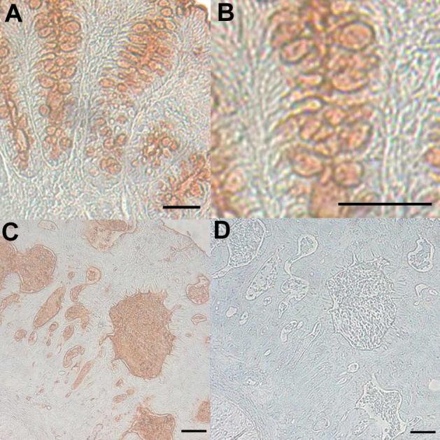
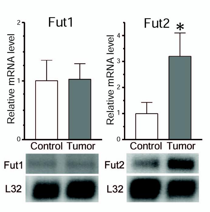
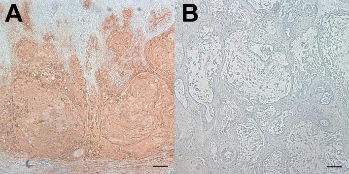
Results: Spontaneous colorectal tumors from Smad3 (-/-) homozygous null mice were found to express alpha(1,2)fucosylated glycans in an abnormal pattern compared to adjacent nonneoplastic colon. Northern blot analysis of alpha(1,2)fucosyltransferase genes Fut1 and Fut2 revealed that Fut2, but not Fut1, steady-state mRNA levels were significantly increased in tumors relative to adjacent normal colonic mucosa. Mutant mice with a Fut2-inactivating germline mutation were crossed with Smad3-targeted mice. In Smad3 (-/-)/Fut2 (-/-) double knockout mice, UEA-I lectin staining was eliminated from colon and colon tumors; however, the number and size of tumors present by 24 weeks of age did not vary regardless of the Fut2 genotype.
Conclusions: In this model of colorectal cancer, cell surface alpha(1,2)fucosylation does not affect development of colon tumors.
Copyright (c) 2007 S. Karger AG, Basel.
Figures
References
-
- Hart GW, Lowe JB, Sathyamoorthy N. Glycobiology and cancer: meeting summary and future diections. Cancer Biol Ther. 2004;3:233–237. - PubMed
-
- Varki A, Marth J. Oligosaccharides in vertebrate development. Seminars Develop Biol. 1995;6:127–138.
-
- Brockhausen I. Pathways of O-glycan biosynthesis in cancer cells. Biochim Biophys Acta. 1999;1473:67–95. - PubMed
-
- Goupille C, Hallouin F, Meflah K, LePendu J. Increase of rat colon carcinoma cells tumorigenicity by alpha(1-2)fucosyltransferase gene transfection. Glycobiology. 1997;7:221–229. - PubMed
Publication types
MeSH terms
Substances
Grants and funding
LinkOut - more resources
Full Text Sources
Molecular Biology Databases
