. 2007 Jan 31;2(1):e177.
doi: 10.1371/journal.pone.0000177.

Selective loss of TGFbeta Smad-dependent signalling prevents cell cycle arrest and promotes invasion in oesophageal adenocarcinoma cell lines

Affiliations

Selective loss of TGFbeta Smad-dependent signalling prevents cell cycle arrest and promotes invasion in oesophageal adenocarcinoma cell lines

Benjamin A Onwuegbusi et al. PLoS One. .

Abstract

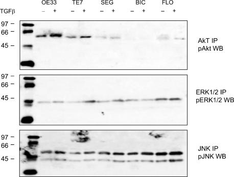
In cancer, Transforming Growth Factor beta (TGFbeta) increases proliferation and promotes invasion via selective loss of signalling pathways. Oesophageal adenocarcinoma arises from Barrett's oesophagus, progresses rapidly and is usually fatal. The contribution of perturbed TGFbeta signalling in the promotion of metastasis in this disease has not been elucidated. We therefore investigated the role of TGFbeta in Barrett's associated oesophageal adenocarcinoma using a panel of cell lines (OE33, TE7, SEG, BIC, FLO). 4/5 adenocarcinoma cell lines failed to cell cycle arrest, down-regulate c-Myc or induce p21 in response to TGFbeta, and modulation of a Smad3/4 specific promoter was inhibited. These hyperproliferative adenocarcinoma cell lines displayed a TGFbeta induced increase in the expression of the extracellular matrix degrading proteinases, urokinase-type plasminogen activator (uPA) and plasminogen activator inhibitor 1 (PAI-1), which correlated with an invasive cell phenotype as measured by in vitro migration, invasion and cell scattering assays. Inhibiting ERK and JNK pathways significantly reduced PAI and uPA induction and inhibited the invasive cell phenotype. These results suggest that TGFbeta Smad-dependent signalling is perturbed in Barrett's carcinogenesis, resulting in failure of growth-arrest. However, TGFbeta can promote PAI and uPA expression and invasion through MAPK pathways. These data would support a dual role for TGFbeta in oesophageal adenocarcinoma.


Conflict of interest statement

Competing Interests: The authors have declared that no competing interests exist.

Figures

Figure 1
Figure 1
Effect of TGFβ on cell cycle progression. All cells were synchronised overnight in serum free media. Cells were then released into cell cycle by complete media (C), or kept continuously in serum free media (SF), or in complete media with TGFβ (10 ng/mL), all for 24 hours. DNA content was then assessed by flow cytometry. (A) Representative FACScan profiles for OE33 and FLO. The initial peak represents the G0/G1 fraction, whilst the second peak represents G2M fraction. (B) Summary of cell cycle distribution for each cell line analysed. Each bar represents mean percentage of total cell population in G0/G1 (grey), S (black) and G2/M (white) phase of the cell cycle from three separate experiments. * p<0.05, *** p<0.001
Figure 2
Figure 2
Regulation of anti-proliferative genes by TGFβ in oesophageal cell lines. mRNA expression of p21 (A) and c-Myc (B) was assessed by quantitative real-time PCR in control cells grown in complete media or in complete media containing 10 ng/mL TGFβ for 24 hours. Results for real-time PCR are expressed as the mean and standard error of four separate experiments relative to β-actin expression. * p<0.05, ***p<0.001
Figure 3
Figure 3
TGFβ stimulation of Smad pathway in oesophageal cell lines. Nuclear translocation (A) and phosphorylation (B) of Smad 2/3 following TGFβ stimulation. Cells were treated with 10 ng/mL of TGFβ1 for 6 h and Smad 2/3 localisation and phosphorylation were determined by immunofluorescence using anti-Smad 2/3 antibody and confocal microscopy and western blotting respectively. Regulation of transcription by TGFβ (C). Cells were co-transfected with the (CAGA)12-Luciferase reporter plasmid and the Renilla Luciferase reporter plasmid then incubated, with or without 10 ng/mL TGFβ for 24 h. Data is expressed as mean fold change in CAGA luciferase activity in TGFβ samples compared to untreated samples, normalised to the activity of Renilla, from four separate experiments * p<0.05, ** p<0.01, ***p<0.001
Figure 4
Figure 4
Regulation of ECM modulating genes by TGFβ. mRNA expression of PAI-1 (A) and uPA (B) was assessed by quantitative real-time PCR in control cells and cells treated with 10 mg/mL TGFβ for 24 hours. Results for real-time PCR expressed as mean and standard error of four separate experiments relative to β-actin expression. * p<0.05, ** p<0.01, ***p<0.001
Figure 5
Figure 5
TGFβ modulation of kinase pathways in BE adenocarcinoma cells. Western blot on lysate from OE33, TE7, SEG, BIC and FLO cells treated with control media (−) or media with 10ng/mL TGFβ (+) for 4 hours for phosphorylated ERK1/2, phosphorylated JNK or phosphorylated Akt. Immunoprecipitation was performed with total antibody for Akt, ERK and JNK, and then blots were probed with the phosphorylated antibody.
Figure 6
Figure 6
Effect of inhibition of PI3K, ERK and JNK pathways on PAI and uPA expression. Quantitative real-time PCR showing PAI (A) and uPA (B) expression in cells treated with 10 ng/mL TGFβ and PI3K inhibitor LY29004 (LY, 20 µM); ERK inhibitor PD98059 (PD, 10 µM); and JNK inhibitor SP600125 (SP, 10 µM) for 24 hours. Results are expressed as a percentage of expression in inhibitor treated samples compared to TGFβ treated samples, such that the greater the inhibition the lower the percentage expression. Results are mean and standard error of four separate experiments. * p<0.05, ** p<0.01, ***p<0.001
Figure 7
Figure 7
Effect of inhibition of PI3K, ERK and JNK pathways on PAI and uPA activity. uPA enzyme activity was assessed by casein zymography, and PAI activity determined by reverse casein zymography in cell lysates from untreated control cells (C), cells treated with 10 ng/mL TGFβ alone (T) and in cells treated with TGFβ and PI3K inhibitor LY294002 (LY), ERK inhibitor PD98059 (PD) or JNK inhibitor SP600125 (SP) for 24 h. uPA activity was detected as digested clear bands on a dark background, whilst PAI activity was detected as dark undigested bands against a clear background.
Figure 8
Figure 8
Effect of inhibition of PI3K, ERK and JNK pathways on invasive properties of oesophageal adenocarcinoma cells following TGFβ stimulation. (A) Aggregation in control cells cultured with normal medium (C), cells treated with TGFβ alone (T) or in the presence of PI3K inhibitor LY294002 (LY), ERK inhibitor PD98590 (PD) or JNK inhibitor SP600125 (SP). Scores represent the mean for 3 separate experiments where 0 is for no aggregates, 1 for small aggregates and 2 for large aggregates. (B) Invasion assay through matrigel matrix over 24 h in untreated cells (C), treated with TGFβ alone (T) or in the presence of inhibitors (LY, PD or SP), (C) Wound healing measured as the percentage of healing of a circular wound over 24 h was assessed in OE33 cells cultured in normal medium (C), or with the addition of TGFβ (T) or with TGFβ in the presence of inhibitors (LY, PD or SP). For all experiments TGFβ is compared with the control and the effect of inhibitors compared with TGFβ. * p<0.05, **p<0.01.

References

    1. Akhurst RJ, Derynck R. TGF-beta signaling in cancer–a double-edged sword. Trends Cell Biol. 2001;11:S44–51. - PubMed
    1. Masui T, Wakefield LM, Lechner JF, LaVeck MA, Sporn MB, et al. Type beta transforming growth factor is the primary differentiation-inducing serum factor for normal human bronchial epithelial cells. Proc Natl Acad Sci U S A. 1986;83:2438–2442. - PMC - PubMed
    1. Liu F, Pouponnot C, Massague J. Dual role of the Smad4/DPC4 tumor suppressor in TGFbeta-inducible transcriptional complexes. Genes Dev. 1997;11:3157–3167. - PMC - PubMed
    1. Bailey T, Biddlestone L, Shepherd N, Barr H, Warner P, et al. Altered cadherin and catenin complexes in the Barrett's esophagus-dysplasia-adenocarcinoma sequence: correlation with disease progression and dedifferentiation. Am J Pathol. 1998;152:135–144. - PMC - PubMed
    1. Seoane J, Pouponnot C, Staller P, Schader M, Eilers M, et al. TGFbeta influences Myc, Miz-1 and Smad to control the CDK inhibitor p15INK4b. Nat Cell Biol. 2001;3:400–408. - PubMed

Publication types

MeSH terms

Substances