. 2007 Jan 31;2(1):e187.
doi: 10.1371/journal.pone.0000187.

Hmgcr in the corpus allatum controls sexual dimorphism of locomotor activity and body size via the insulin pathway in Drosophila

Affiliations

Hmgcr in the corpus allatum controls sexual dimorphism of locomotor activity and body size via the insulin pathway in Drosophila

Yesser Hadj Belgacem et al. PLoS One. .

Abstract

The insulin signaling pathway has been implicated in several physiological and developmental processes. In mammals, it controls expression of 3-Hydroxy-3-Methylglutaryl CoA Reductase (HMGCR), a key enzyme in cholesterol biosynthesis. In insects, which can not synthesize cholesterol de novo, the HMGCR is implicated in the biosynthesis of juvenile hormone (JH). However, the link between the insulin pathway and JH has not been established. In Drosophila, mutations in the insulin receptor (InR) decrease the rate of JH synthesis. It is also known that both the insulin pathway and JH play a role in the control of sexual dimorphism in locomotor activity. In studies here, to demonstrate that the insulin pathway and HMGCR are functionally linked in Drosophila, we first show that hmgcr mutation also disrupts the sexual dimorphism. Similarly to the InR, HMGCR is expressed in the corpus allatum (ca), which is the gland where JH biosynthesis occurs. Two p[hmgcr-GAL4] lines were therefore generated where RNAi was targeted specifically against the HMGCR or the InR in the ca. We found that RNAi-HMGCR blocked HMGCR expression, while the RNAi-InR blocked both InR and HMGCR expression. Each RNAi caused disruption of sexual dimorphism and produced dwarf flies at specific rearing temperatures. These results provide evidence: (i) that HMGCR expression is controlled by the InR and (ii) that InR and HMGCR specifically in the ca, are involved in the control of body size and sexual dimorphism of locomotor activity.


Conflict of interest statement

Competing Interests: The authors have declared that no competing interests exist.

Figures

Figure 1
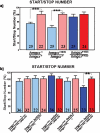
Figure 1. The sexual dimorphism in locomotor activity is disrupted in hmgcr mutant.
a) Flies homozygous for a P element inserted in the 3′ region of the hmgcr gene (hmgcr11635/hmgcr11635) do not exhibit a sexually dimorphic start/stop number. Flies heterozygous for this P element insertion (hmgcr11635/CS) behave like wild-type controls Canton-S flies. For all graphics representing the start/stop number in this study, blue and red are males and females respectively, while the number in the boxes indicates the number of flies recorded for each genotype. CS = Canton-S. Mean±SEM is represented and all statistical tests are done using an Anova-Manova test (Statistica software). * p<0.05; ** p<0.01; *** p<0.001. b) Targeted expression of p[UAS-clb] under the control the p[da-GAL4] driver in flies homozygous for the hmgcr11635 allele (p[da-GAL4]; hmgcr11635/UAS-clb, hmgcr11635) is sufficient to rescue both the lethality and the sexual dimorphism, observed in hmgcr11635/hmgcr11635 flies. However, the p[act-GAL4] driver (p[act-GAL4]; hmgcr11635/UAS-clb, hmgcr11635) rescue the lethality, but not the sexual dimorphism.
Figure 2
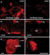
Figure 2. hmgcr is expressed in the brain, the larval fat body, the cardia and the corpus allatum.
Immunohistological staining showing the HMGCR expression in various tissues, in adult fly. a, b, c) The HMGCR is expressed in the fat body in flies younger than 4 days (a, left). In flies older than 5 days, no HMGCR expression is detectable (a, right). The HMGCR is also expressed in some neurons, principally in the cortex of the brain (cx) (b) and some cells of the pars intercerebralis (PI) (c). oe: eosophagus, eb: ellipsoid-body. d, e) In the body, the reductase is detected in the cardia (d) and in the corpus allatum (ca) (e). For a negative control of the HMGCR staining, see Figure 5b, in comparison with figures 5a, c, d, e and g. Scale bar = 25 µm.
Figure 3
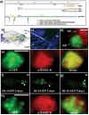
Figure 3. Genesis and expression pattern of the two transgenic p[hmgcr-GAL4] lines.
a) Promoters and genomic regions of hmgcr gene. Two mRNAs are transcribed (mRNAA and mRNAB) under the control of two distinct promoter regions. After splicing, the encoding regions of the two cDNA are identical. A computer analysis of the genomic DNA region has allowed to find two E-Box consensus sequences (CATGTG) localized at −3194 to −3188 and −1315 to −1309, and one E-Box High affinity (GATGACCCGGTCGAGGAG) localized at −2051 to −2033, comprised in a region of 3,8 kb just upstream of the first exon of hmgcr. We then hypothesized that this region could control the RNAA transcription, and cloned and used it to generate the p[DI-3-GAL4] (DI-3) line. To generate the p[DI-11-GAL4] (DI-11) line, we cloned a fragment of 10,6 kb of the first intron (+2733 to +13525) in which we found an E-box consensus (+2911 to +2916) and one SRE1 (AATTAGTCTGTACCCCAATT) (+3551 to +3570). We hypothesized that this region could control the RNAB transcription. b) Schematic lateral view of the head and thorax of Drosophila. card: cardia; ca: corpus allatum; br: brain. c–h) The two p[DI-GAL4] lines (DI-3 and DI-11) drive the expression of GFP in the ca. c) At low magnification (10X), the ca, in green, is detected by using primary antibodies against GFP and secondary antibodies labeled with FITC (green) in flies DI-3/GFP. The background is artificially colored in blue. d) The corpus cardiacum (cc) is a structure located near the ca. To confirm that the GFP is expressed in the ca, the corpus cardiacum (cc) was stained using an anti-AKH antibody directly conjugated to the rhodamine (red). Thus, we can visualize the ca in green and the cc in red. e) The GFP (left, green) and HMGCR (middle, red) are detected with primary and secondary antibody. Those last are labeled respectively with FITC (green) or Cy3 (red). When merged, we can see that the GFP and HMGCR are colocalized (right panel: yellow), confirming that the DI-3 drives the GFP in the same cells of the ca that express the HMGCR. f) Dissection of the cardia and the ca from DI-3/UAS-gfp flies of different ages and observed directly under a binocular lamp fit with a green fluorescent filter (Leica, MZ FLIII). The GFP is driven by DI-3 in the ca in a temporal dynamic pattern. In two days old adult flies (left panel), the GFP is well detectable in the ca (arrowhead), but not anymore in 5 days old flies (right panel). However, in 5 days old flies, the GFP is detectable in a thin layer of the cardia. g) Dissection of cardia and ca from (DI-11/UAS-gfp) directly observed under a binocular lamp fit with a green fluorescent filter (Leica, MZ FLIII). DI-11 also drives the expression of GFP in the ca (arrowhead), and this expression is permanent in adult flies. h) GFP (left) and HMGCR (middle) are detected respectively with primary antibodies against GFP and HMGCR and revealed with secondary antibodies respectively labeled with FITC (green) and Cy3 (red). When merged, we can see that the GFP and HMGCR are colocalized (right panel: yellow), confirming that the DI-11 drives the GFP in the same cells of the ca that express the HMGCR. Scale bar = 25 µm.
Figure 4
Figure 4. Targeted expression of the HMGCR in the corpus allatum rescues the hmgcr mutant phenotype.
p[DI-3-GAL4] plus p[act-GAL4] in the same flies are sufficient to rescue the sexual dimorphism disrupted in hmgcr11635/hmgcr11635 flies (DI-3, act-GAL4; hmgcr11635/UAS-clb; hmgcr11635). This suggests a specific role for the HMGCR in the ca to control the sexual dimorphism. Interestingly, this rescue occurs in two days old flies, but not in the same flies of 5 days old.
Figure 5
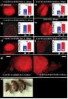
Figure 5. Directed expression of p[UAS-RNAi-HMGCR] specifically in the corpus allatum mimics the hmgcr mutation, by disrupting the sexual dimorphism.
For all panels (a–g): immunostaining using a primary antibody raised against the human form of the HMGCR, revealed with a secondary antibody labeled with Cy3 (red). a–d) Targeting UAS-RNAi-HMGCR specifically in the ca yields to the lack of the HMGCR protein, and leads to the disruption of the sexual dimorphism. This effect is completely reversible. In 2 days old DI-3/UAS-RNAi-HMGCR flies (b), HMGCR protein is not detectable and the number of start/stop is identical between males and females compared to controls (a) (2 days old UAS-RNAi-HMGCR/CS flies). Interestingly, in 5 days old flies, both DI-3/UAS-RNAi-HMGCR (d) and UAS-RNAi-HMGCR/CS (c) the HMGCR is detectable and the start/stop number is sexually dimorphic. This is in agreement with the temporal expression pattern driven by the DI-3. Additionally, this result strongly suggests the reversibility of the RNAi effect. N.B: results from b) and d) come from the same flies, recorded at 2 and 5 days old respectively. e, f) Expressing the UAS-RNAi-HMGCR in the ca using the DI-11 line (DI-11/UAS-RNAi-HMGCR) also leads to the lack of the HMGCR product (f) and disrupts the sexual dimorphism compared to controls flies (e). g) The HMGCR is expressed both in the cardia and the ca (left panel) in control flies (2 days old UAS-RNAi-HMGCR/CS flies), whereas in 2 days old (DI-3/UAS-RNAi-HMGCR) flies, the HMGCR is detected only in the cardia (right panel). This last staining serves as a positive control, to demonstrate that the DI-3 drives the UAS-RNAi-HMGCR only in the ca. h) Expressing the UAS-RNAi-HMGCR in the ca under the control of DI-11 leads to a strong lethality when flies are reared at 24°C. Moreover, the only few females that survived are dwarf (left: dwarf female DI-11/UAS-RNAi-HMGCR, middle: female control UAS-RNAi-HMGCR/CS] and right: female CS). Scale bar = 25 µm.
Figure 6
Figure 6. Directed expression of p[UAS-RNAi-InR] specifically in the corpus allatum, mimics the hmgcr mutation.
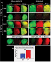
For all panels (a–c): immunostaining using a primary antibody raised against the human form of the InR, revealed by a secondary antibody labeled with FITC (green). a–c) Targeting the UAS-RNAi-InR specifically in the ca blocks the InR expression and disrupts the sexual dimorphism. This effect is completely reversible. In 2 days old DI-3/UAS-RNAi-InR flies (b), InR is not detectable and the number of start/stop is identical between males and females compared to controls flies (a) (2 days old UAS-RNAi-InR/CS). Interestingly, in 5 days old DI-3/UAS-RNAi-InR flies (c) the InR is detectable and the start/stop number is sexually dimorphic. This result corroborates the temporal expression pattern driven by the DI-3. Additionally, this strongly suggests, like for the RNAi-HMGCR, the reversibility of the RNAi-InR effect. N.B.: again here, results from b) and d) come from the same flies, recorded at 2 and 5 days old, respectively. d) Expressing the UAS-RNAi-InR in the ca under the control of DI-11 leads to a strong lethality when flies are reared at 24°C and 19°C. However, at 19°C, few females survive, but they are dwarf (left: dwarf female DI-11/UAS-RNAi-InR, right: female control (UAS-RNAi-InR/CS). Scale bar = 25 µm.
Figure 7
Figure 7. Insulin signaling pathway controls the sexual dimorphism via HMGCR.
a) Immunostaining using primary antibodies against the InR and/or HMGCR, revealed by secondary antibodies labeled with FITC (green) or Cy3 (red) respectively. HMGCR and InR are colocalized in the same cells of the ca. Blocking the expression of the HMGCR does not influence the expression of the InR, but abolishes the sexual dimorphism. However, blocking the expression of the InR blocks the expression of the HMGCR and consequently abolishes the sexual dimorphism. This suggests that the InR controls the expression of the HMGCR within the ca. To test this hypothesis, we directed the expression of the UAS-clb concomitantly with the UAS-RNAi-InR (DI-3/UAS-RNAi-InR; UAS-clb). In this case, although InR is not detectable, HMGCR is present in the ca and the start/stop number is sexually dimorphic between males and females (b). Scale bar = 25 µm.
Figure 8
Figure 8. Model of the insulin pathway regulating the expression of the HMGCR in the corpus allatum.
We hypothesize that the two different populations of cells, the Insulin Producing Cells (IPCs) and the feminising cells (FCs), located in the pars intercerebralis control, by two distinct ways, the JH synthesis/release by the corpus allatum. The informative way (in yellow), which could be feminised, might control the level or the timing of the JH release, while the permissive way (in green) might control or allow the presence of the molecular machinery, as the transcription of the HMGCR, to promote the JH synthesis. In magnification: in the corpus allatum, insulin binds to its receptor (InR), which activates SREBP. In turn, SREBP regulates the transcription of hmgcr gene.

References

    1. Takahashi Y, Tobe K, Kadowaki H, Katsumata D, Fukushima Y, et al. Roles of insulin receptor substrate-1 and Shc on insulin-like growth factor I receptor signaling in early passages of cultured human fibroblasts. Endocrinology. 1997;138:741–750. - PubMed
    1. Butler AA, Le Roith D. Control of growth by the somatropic axis: growth hormone and the insulin-like growth factors have related and independent roles. Annu Rev Physiol. 2001;63:141–164. - PubMed
    1. Saltiel AR, Kahn CR. Insulin signalling and the regulation of glucose and lipid metabolism. Nature. 2001;414:799–806. - PubMed
    1. Foufelle F, Ferre P. [Regulation of carbohydrate metabolism by insulin: role of transcription factor SREBP-1c in the hepatic transcriptional effects of the hormone]. J Soc Biol. 2001;195:243–248. - PubMed
    1. Penhos JC, Wu CH, Lemberg A, Daunas J, Brodoff B, et al. The effect of insulin on the metabolism of lipids and on urea formation by the perfused rat liver. Metabolism. 1968;17:246–259. - PubMed

Publication types

MeSH terms