

Diversity of sympathetic vasoconstrictor pathways and their plasticity after spinal cord injury
- PMID: 17264977
- PMCID: PMC1797061
- DOI: 10.1007/s10286-006-0394-8
Diversity of sympathetic vasoconstrictor pathways and their plasticity after spinal cord injury
Abstract
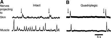
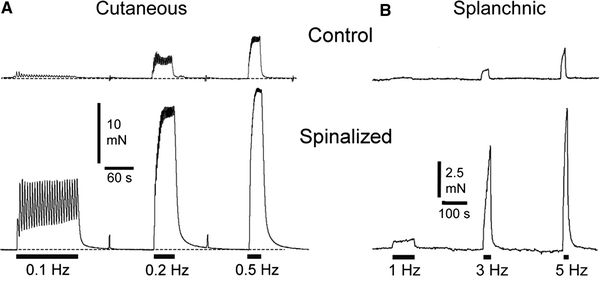
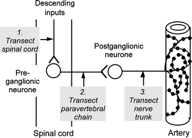
Sympathetic vasoconstrictor pathways pass through paravertebral ganglia carrying ongoing and reflex activity arising within the central nervous system to their vascular targets. The pattern of reflex activity is selective for particular vascular beds and appropriate for the physiological outcome (vasoconstriction or vasodilation). The preganglionic signals are distributed to most postganglionic neurones in ganglia via synapses that are always suprathreshold for action potential initiation (like skeletal neuromuscular junctions). Most postganglionic neurones receive only one of these "strong" inputs, other preganglionic connections being ineffective. Pre- and postganglionic neurones discharge normally at frequencies of 0.5-1 Hz and maximally in short bursts at <10 Hz. Animal experiments have revealed unexpected changes in these pathways following spinal cord injury. (1) After destruction of preganglionic neurones or axons, surviving terminals in ganglia sprout and rapidly re-establish strong connections, probably even to inappropriate postganglionic neurones. This could explain aberrant reflexes after spinal cord injury. (2) Cutaneous (tail) and splanchnic (mesenteric) arteries taken from below a spinal transection show dramatically enhanced responses in vitro to norepinephrine released from perivascular nerves. However the mechanisms that are modified differ between the two vessels, being mostly postjunctional in the tail artery and mostly prejunctional in the mesenteric artery. The changes are mimicked when postganglionic neurones are silenced by removal of their preganglionic input. Whether or not other arteries are also hyperresponsive to reflex activation, these observations suggest that the greatest contribution to raised peripheral resistance in autonomic dysreflexia follows the modifications of neurovascular transmission.
Figures
Similar articles
-
Adaptations of peripheral vasoconstrictor pathways after spinal cord injury.Prog Brain Res. 2006;152:289-97. doi: 10.1016/S0079-6123(05)52019-3. Prog Brain Res. 2006. PMID: 16198708 Review.
-
Changes in sympathetic neurovascular function following spinal cord injury.Auton Neurosci. 2018 Jan;209:25-36. doi: 10.1016/j.autneu.2017.02.003. Epub 2017 Feb 11. Auton Neurosci. 2018. PMID: 28209424 Review.
-
Transmission of signals through sympathetic ganglia--modulation, integration or simply distribution?Acta Physiol Scand. 2003 Mar;177(3):227-35. doi: 10.1046/j.1365-201X.2003.01075.x. Acta Physiol Scand. 2003. PMID: 12608993 Review.
-
Effects of spinal cord injury on synaptic inputs to sympathetic preganglionic neurons.Prog Brain Res. 2006;152:11-26. doi: 10.1016/S0079-6123(05)52001-6. Prog Brain Res. 2006. PMID: 16198690 Review.
-
Reflex activation of postganglionic vasoconstrictor neurones supplying skeletal muscle by stimulation of arterial chemoreceptors via non-nicotinic synaptic mechanisms in sympathetic ganglia.Pflugers Arch. 1983 Feb;396(2):95-100. doi: 10.1007/BF00615511. Pflugers Arch. 1983. PMID: 6835822
Cited by
-
Somatosympathetic Vasoconstrictor Reflexes in Human Spinal Cord Injury: Responses to Innocuous and Noxious Sensory Stimulation below Lesion.Front Physiol. 2012 Jun 25;3:215. doi: 10.3389/fphys.2012.00215. eCollection 2012. Front Physiol. 2012. PMID: 22737131 Free PMC article.
-
Dramatically Amplified Thoracic Sympathetic Postganglionic Excitability and Integrative Capacity Revealed with Whole-Cell Patch-Clamp Recordings.eNeuro. 2019 May 13;6(2):ENEURO.0433-18.2019. doi: 10.1523/ENEURO.0433-18.2019. Print 2019 Mar/Apr. eNeuro. 2019. PMID: 31040159 Free PMC article.
-
A systematic review of the management of autonomic dysreflexia after spinal cord injury.Arch Phys Med Rehabil. 2009 Apr;90(4):682-95. doi: 10.1016/j.apmr.2008.10.017. Arch Phys Med Rehabil. 2009. PMID: 19345787 Free PMC article.
-
Orgasm and SCI: what do we know?Spinal Cord. 2018 Jun;56(6):538-547. doi: 10.1038/s41393-017-0020-8. Epub 2017 Dec 20. Spinal Cord. 2018. PMID: 29259346 Review.
-
Skin vasomotor hemiparesis followed by overactivity: characteristic thermography findings in a patient with Horner syndrome due to spinal cord infarction.Clin Auton Res. 2016 Apr;26(2):153-6. doi: 10.1007/s10286-016-0344-z. Epub 2016 Jan 25. Clin Auton Res. 2016. PMID: 26811085
References
-
- {'text': '', 'ref_index': 1, 'ids': [{'type': 'DOI', 'value': '10.1002/cne.902830208', 'is_inner': False, 'url': 'https://doi.org/10.1002/cne.902830208'}, {'type': 'PubMed', 'value': '2567744', 'is_inner': True, 'url': 'https://pubmed.ncbi.nlm.nih.gov/2567744/'}]}
- Anderson CR, McLachlan EM, Srb-Christie O (1989) Distribution of sympathetic preganglionic neurons and monoaminergic nerve terminals in the spinal cord of the rat. J Comp Neurol 283:269–284 - PubMed
-
- {'text': '', 'ref_index': 1, 'ids': [{'type': 'DOI', 'value': '10.1152/ajpheart.00712.2005', 'is_inner': False, 'url': 'https://doi.org/10.1152/ajpheart.00712.2005'}, {'type': 'PubMed', 'value': '16143650', 'is_inner': True, 'url': 'https://pubmed.ncbi.nlm.nih.gov/16143650/'}]}
- Brock JA, Yeoh M, McLachlan EM (2006) Enhanced neurally evoked responses and inhibition of norepinephrine reuptake in rat mesenteric arteries after spinal transection. Am J Physiol––Heart Circ Physiol 290:H398–405 - PubMed
-
- {'text': '', 'ref_index': 1, 'ids': [{'type': 'DOI', 'value': '10.1046/j.1440-1681.2002.03640.x', 'is_inner': False, 'url': 'https://doi.org/10.1046/j.1440-1681.2002.03640.x'}, {'type': 'PubMed', 'value': '11985533', 'is_inner': True, 'url': 'https://pubmed.ncbi.nlm.nih.gov/11985533/'}]}
- Dampney RA, Coleman MJ, Fontes MA, Hirooka Y, Horiuchi J, Li YW, Polson JW, Potts PD, Tagawa T (2002) Central mechanisms underlying short- and long-term regulation of the cardiovascular system. Clin Exp Pharmacol Physiol 29:261–268 - PubMed
-
- {'text': '', 'ref_index': 1, 'ids': [{'type': 'PubMed', 'value': '7735278', 'is_inner': True, 'url': 'https://pubmed.ncbi.nlm.nih.gov/7735278/'}]}
- Dembowsky K (1995) Integrative properties of sympathetic preganglionic neurones within the thoracic spinal cord. Clin Exp Hypertens 17:313–321 - PubMed
-
- {'text': '', 'ref_index': 1, 'ids': [{'type': 'DOI', 'value': '10.1016/0165-1838(85)90012-8', 'is_inner': False, 'url': 'https://doi.org/10.1016/0165-1838(85)90012-8'}, {'type': 'PubMed', 'value': '2993402', 'is_inner': True, 'url': 'https://pubmed.ncbi.nlm.nih.gov/2993402/'}]}
- Dembowsky K, Czachurski J, Seller H (1985) An intracellular study of the synaptic input to sympathetic preganglionic neurones of the third thoracic segment of the cat. J Auton Nerv Syst 13:201–244 - PubMed
Publication types
MeSH terms
LinkOut - more resources
Full Text Sources
Medical
