

AVPV neurons containing estrogen receptor-beta in adult male rats are influenced by soy isoflavones
- PMID: 17266774
- PMCID: PMC1797051
- DOI: 10.1186/1471-2202-8-13
AVPV neurons containing estrogen receptor-beta in adult male rats are influenced by soy isoflavones
Abstract
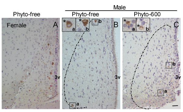
Background: Isoflavones, the most abundant phytoestrogens in soy foods, are structurally similar to 17beta-estradiol. It is known that 17beta-estradiol induces apoptosis in anteroventral periventricular nucleus (AVPV) in rat brain. Also, there is evidence that consumption of soy isoflavones reduces the volume of AVPV in male rats. Therefore, in this study, we examined the influence of dietary soy isoflavones on apoptosis in AVPV of 150 day-old male rats fed either a soy isoflavone-free diet (Phyto-free) or a soy isoflavone-rich diet (Phyto-600).
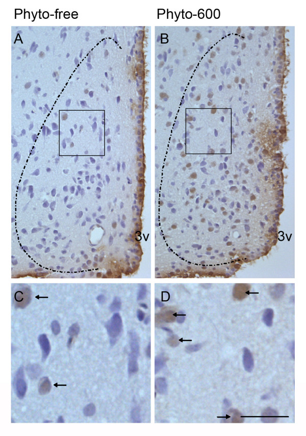
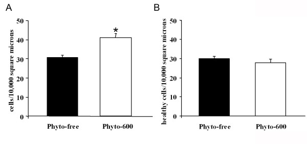
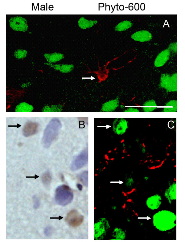
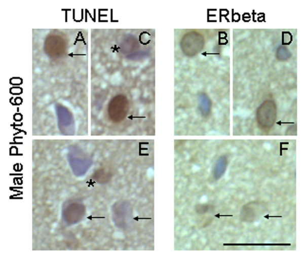
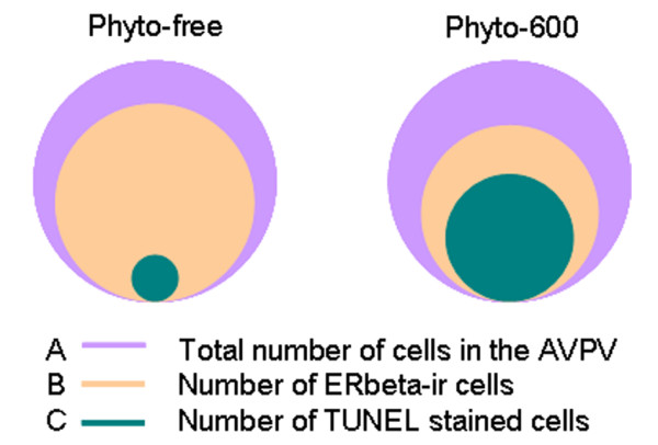
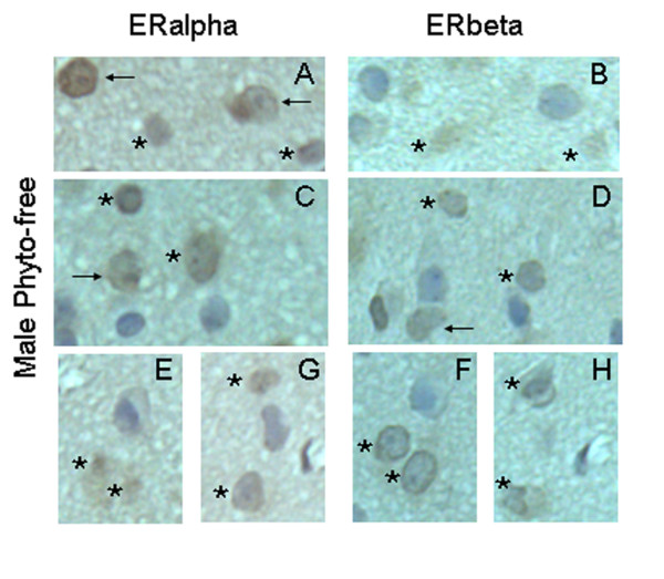
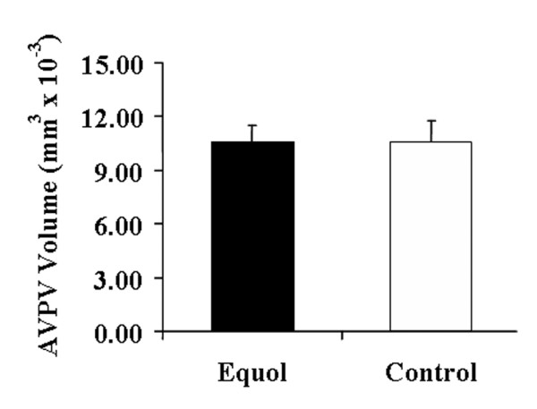
Results: The occurrence of apoptosis in AVPV was examined by TUNEL staining. The incidence of apoptosis was about 10 times higher in the Phyto-600 group (33.1 +/- 1.7%) than in the Phyto-free group (3.6 +/- 1.0%). Furthermore, these apoptotic cells were identified as neurons by dual immunofluorescent staining of GFAP and NeuN as markers of astrocytes and neurons, respectively. Then the dopaminergic neurons in AVPV were detected by immunohistochemistry staining of tyrosine hydroxylase (TH). No significant difference in the number of TH neurons was observed between the diet treatment groups. When estrogen receptor (ER) alpha and beta were examined by immunohistochemistry, we observed a 22% reduction of ERbeta-positive cell numbers in AVPV with consumption of soy isoflavones, whereas no significant change in ERalpha-positive cell numbers was detected. Furthermore, almost all the apoptotic cells were ERbeta-immunoreactive (ir), but not ERalpha-ir. Last, subcutaneous injections of equol (a major isoflavone metabolite) that accounts for approximately 70-90% of the total circulating plasma isoflavone levels did not alter the volume of AVPV in adult male rats.
Conclusion: In summary, these findings provide direct evidence that consumption of soy isoflavones, but not the exposure to equol, influences the loss of ERbeta-containing neurons in male AVPV.
Figures

Similar articles
-
Soy isoflavones modulate the expression of BAD and neuron-specific beta III tubulin in male rat brain.Neurosci Lett. 2005 Sep 9;385(2):153-7. doi: 10.1016/j.neulet.2005.05.040. Neurosci Lett. 2005. PMID: 15951108
-
Isoflavones made simple - genistein's agonist activity for the beta-type estrogen receptor mediates their health benefits.Med Hypotheses. 2006;66(6):1093-114. doi: 10.1016/j.mehy.2004.11.046. Epub 2006 Mar 2. Med Hypotheses. 2006. PMID: 16513288
-
Estrogens and phytoestrogens: brain plasticity of sexually dimorphic brain volumes.J Steroid Biochem Mol Biol. 2003 Jun;85(2-5):299-309. doi: 10.1016/s0960-0760(03)00210-3. J Steroid Biochem Mol Biol. 2003. PMID: 12943716
-
Neurobehavioral effects of dietary soy phytoestrogens.Neurotoxicol Teratol. 2002 Jan-Feb;24(1):5-16. doi: 10.1016/s0892-0362(01)00197-0. Neurotoxicol Teratol. 2002. PMID: 11836067 Review.
-
Early life and adult exposure to isoflavones and breast cancer risk.J Environ Sci Health C Environ Carcinog Ecotoxicol Rev. 2008 Apr-Jun;26(2):113-73. doi: 10.1080/10590500802074256. J Environ Sci Health C Environ Carcinog Ecotoxicol Rev. 2008. PMID: 18569328 Review.
Cited by
-
Dietary phytoestrogens recalibrate socioemotional behavior in C57Bl/6J mice in a sex- and timing-dependent manner.Horm Behav. 2025 Feb;168:105678. doi: 10.1016/j.yhbeh.2025.105678. Epub 2025 Jan 17. Horm Behav. 2025. PMID: 39826371
-
Estrogenic environmental endocrine-disrupting chemical effects on reproductive neuroendocrine function and dysfunction across the life cycle.Rev Endocr Metab Disord. 2007 Jun;8(2):143-59. doi: 10.1007/s11154-007-9048-y. Rev Endocr Metab Disord. 2007. PMID: 17674209 Review.
-
Sexual dimorphism in the hypophysiotropic tyrosine hydroxylase-positive neurons in the preoptic area of the teleost, Clarias batrachus.Biol Sex Differ. 2015 Nov 9;6:23. doi: 10.1186/s13293-015-0042-x. eCollection 2015. Biol Sex Differ. 2015. PMID: 26557978 Free PMC article.
-
Sexual differentiation of the brain requires perinatal kisspeptin-GnRH neuron signaling.J Neurosci. 2014 Nov 12;34(46):15297-305. doi: 10.1523/JNEUROSCI.3061-14.2014. J Neurosci. 2014. PMID: 25392497 Free PMC article.
-
Comparative study of environmental pollutants bisphenol A and bisphenol S on sexual differentiation of anteroventral periventricular nucleus and spermatogenesis.Reprod Biol Endocrinol. 2019 Jul 10;17(1):53. doi: 10.1186/s12958-019-0491-x. Reprod Biol Endocrinol. 2019. PMID: 31292004 Free PMC article.
References
-
- Knight DC, Eden JA. A review of the clinical effects of phytoestrogens. Obstet Gynecol. 1996;87:897–904. - PubMed
-
- Setchell KD, Brown NM, Lydeking-Olsen E. The clinical importance of the metabolite equol-a clue to the effectiveness of soy and its isoflavones. J Nutr. 2002;132:3577–3584. - PubMed
-
- Setchell KD, Clerici C, Lephart ED, Cole SJ, Heenan C, Castellani D, Wolfe BE, Nechemias-Zimmer L, Brown NM, Lund TD, Handa RJ, Heubi JE. S-equol, a potent ligand for estrogen receptor beta, is the exclusive enantiomeric form of the soy isoflavone metabolite produced by human intestinal bacterial flora. Am J Clin Nutr. 2005;81:1072–1079. - PubMed
Publication types
MeSH terms
Substances
LinkOut - more resources
Full Text Sources
Miscellaneous
