. 2007;35(4):1289-300.
doi: 10.1093/nar/gkm016. Epub 2007 Jan 31.

Identification and characterization of human Mex-3 proteins, a novel family of evolutionarily conserved RNA-binding proteins differentially localized to processing bodies

Affiliations

Identification and characterization of human Mex-3 proteins, a novel family of evolutionarily conserved RNA-binding proteins differentially localized to processing bodies

Karine Buchet-Poyau et al. Nucleic Acids Res. 2007.

Abstract

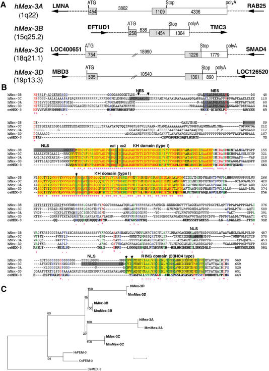
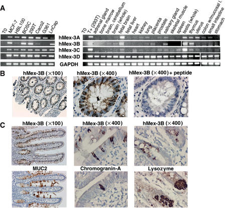
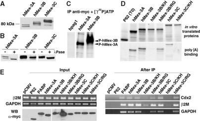
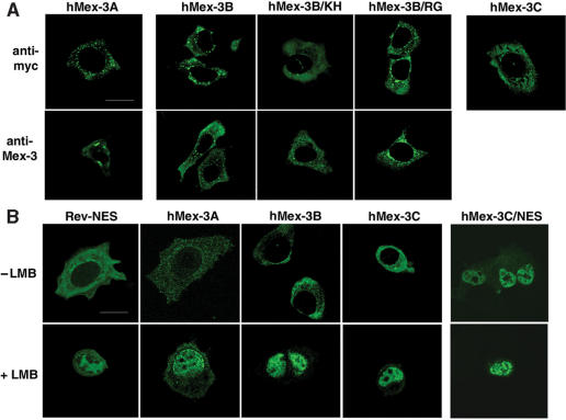
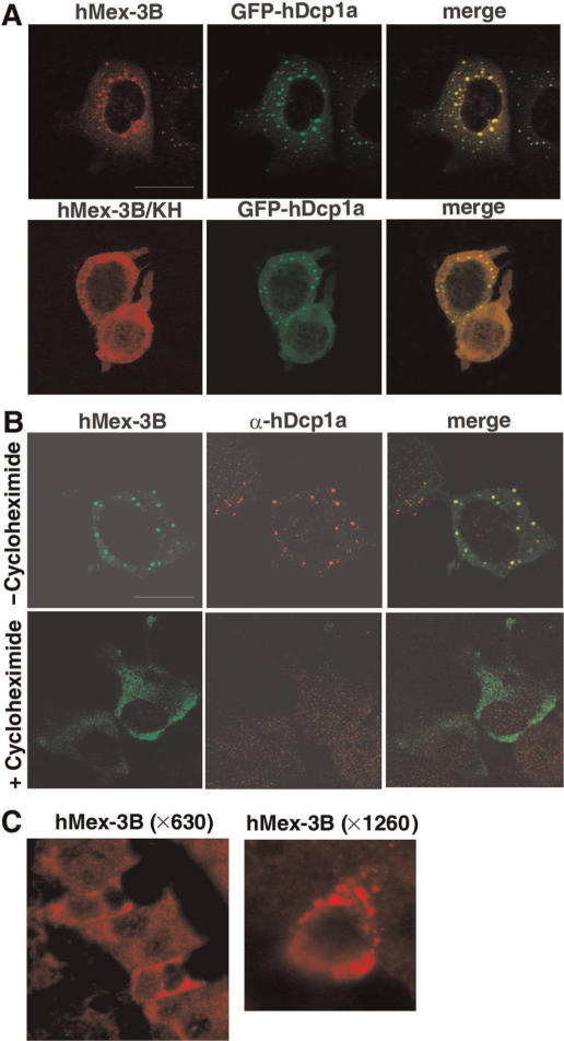
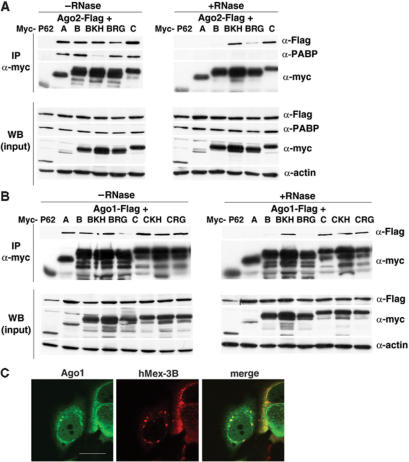
In Caenorhabditis elegans, the Mex-3 protein is a translational regulator that specifies the posterior blastomere identity in the early embryo and contributes to the maintenance of the germline totipotency. We have now identified a family of four homologous human Mex-3 genes, called hMex-3A to -3D that encode proteins containing two heterogeneous nuclear ribonucleoprotein K homology (KH) domains and one carboxy-terminal RING finger module. The hMex-3 are phosphoproteins that bind RNA through their KH domains and shuttle between the nucleus and the cytoplasm via the CRM1-dependent export pathway. Our analysis further revealed that hMex-3A and hMex-3B, but not hMex-3C, colocalize with both the hDcp1a decapping factor and Argonaute (Ago) proteins in processing bodies (P bodies), recently characterized as centers of mRNA turnover. Taken together, these findings indicate that hMex-3 proteins constitute a novel family of evolutionarily conserved RNA-binding proteins, differentially recruited to P bodies and potentially involved in post-transcriptional regulatory mechanisms.


Figures

Figure 1.
Figure 1.
Human family of Mex-3 proteins. (A) Structure and chromosomal localization of the four human Mex-3 genes. Exons are depicted as boxes, with coding sequences as grey boxes and 3’-UTR as white boxes. Genes lying downstream and upstream of hMex-3A, hMex-3B, hMex-3C and hMex-3D genes are displayed as large arrows. (B) Sequence alignment of the hMex-3 proteins and their Caenorhabditis elegans ortholog. C. elegans Mex-3 and hMex-3 amino acid sequences were aligned with ClustalW. Identical residues (red) are annotated by an asterisk, whereas similar residues (green) and lightly similar residues (blue) are denoted with two or one dot, respectively. Junctions between the two exons (vertical line) lie in the same position for the four hMex-3 mRNA. One NES is predicted in each hMex-3 protein, and one NLS is also predicted for hMex-3B and hMex-3C. Two strongly conserved KH domains of type I were predicted in hMex-3 proteins. One conserved RING domain of C3H4 type was also predicted in C-terminus of the four human proteins. The highly conserved amino acids of KH and RING domains are shown as green boxes. Arrows indicate the amino acids which were mutated in this study. (C) Phylogenetic tree of the Mex-3 gene family. The 180 most conserved amino acids around the KH domains of human Mex-3, mouse Mex-3, ceMex-3 (Genbank accession number AAK73873) and two Ascidian Mex-3 homologous sequences from Ciona savignyi and Halocynthia roretzi (Genbank accession number BAB03404 and BAC10968) were aligned by T-coffee. A tree was done with PhyML method on this alignment. Bootstrap values are indicated as percentages (500 replicates). Scale bar: 0.05.
Figure 2.
Figure 2.
Expression profile of hMex-3 genes and hMex-3B protein. (A) Human Mex-3 gene expression levels (panels 1–4) were examined by RT-PCR with specific internal primers and were compared with expression level of ubiquitously expressed GAPDH gene (panel 5). RNA were extracted from 7 human cell lines (left) and from 20 human tissues (Multiple Tissue Total RNA panel, BD Biosciences) (right). (B) Human colon sections (magnification: ×100 or ×400) were stained with anti-hMex-3Bβ antibody (panels 1 and 2), anti-hMex-3Bβ antibody + hMex-3B peptide, as control (panel 3). (C) Serial sections of human Meckel's diverticulum (magnification: ×100 or ×400) were stained with anti-hMex-3Bβ (top left) and anti-MUC2 (bottom left), anti-hMex-3Bβ (top middle) and anti-Chromogranin-A (bottom middle), anti-hMex-3Bβ (top right) and anti-Lysozyme (bottom right).
Figure 3.
Figure 3.
Biochemical characterization of hMex-3 proteins. (A and B) BOSC cells were transiently transfected with vectors expressing myc-tagged forms of hMex-3A, -3B and -3C. Western blot analysis was performed with anti-myc antibody. In (B) treatment of protein extracts with (+) or without (−) λ-Phosphatase. (C) Kinase assay. hMex-3A and -3B proteins or a control protein (Bpag1) expressed in BOSC cells were immunoprecipitated with the anti-myc antibody and incubated with kinase buffer and [γ32P] ATP. Labeled proteins were revealed by autoradiography. (D) RNA homopolymer binding assay. Proteins from indicated expression vectors were in vitro translated in the presence of [35S] methionine (top). Binding to agarose beads coupled to poly(A) (bottom) RNA homopolymers is shown for in vitro translated proteins. As a negative control, a fragment of P62-sequestosome protein was incubated with RNA homopolymers in the same conditions. One-tenth of the initial translation reactions and all the bound proteins were analysed by SDS-PAGE and autoradiography. (E) In vivo hMex-3 binding to mRNA. BOSC cells were transiently transfected with vectors expressing myc-tagged proteins, as indicated. RT-PCR amplification was performed on total RNA extracted from those cells (top left). Western blot analysis performed with anti-myc antibody (bottom left). RT-PCR amplification performed on total RNA extracted from sepharose-protein A beads after immunoprecipitation by an anti-myc antibody (right).
Figure 4.
Figure 4.
Subcellular localization of hMex-3 proteins. (A) MCF7 cells expressing hMex-3A, -3B and -3C were stained with anti-myc antibody (top) or with specific anti-hM3Aβ or anti-hM3Bβ antibodies (bottom) and revealed by FITC-conjugated secondary antibodies. (B) MCF7 cells expressing hMex-3A, -3B, -3C proteins or hMex-3C mutated within the nuclear export signal (NES) sequence were treated with or without 20 ng/ml of Leptomycin B (LMB) and were stained as above. pEGFP-Rev-NES construct was used as a positive control. Scale bar: 20 μm.
Figure 5.
Figure 5.
Localization of hMex-3B in Dcp-containing bodies. (A) MCF7 cells expressing hMex-3B or hMex-3B/KH and GFP-hDcp1a proteins were stained with anti-hM3Bα antibody and revealed with ALEXA555-conjugated secondary antibody (left). Central panel shows GFP signal. Right panel shows overlay of the two signals. (B) MCF7 cells expressing hMex-3B protein treated with or without 5 μg/ml of cycloheximide were stained with anti-myc antibody (left) and anti-hDcp1a antibody (middle) and revealed with ALEXA488 and ALEXA555-conjugated secondary antibodies. Right panels show overlay of the two signals. Scale bar: 20 μm. (C) Sections of mouse duodenum (magnification: ×630 or ×1260) were stained with anti-hMex-3Bβ and revealed with an ALEXA555-conjugated secondary antibody. Tissues were mounted and observed by a confocal microscope, as described in the materials and methods section.
Figure 6.
Figure 6.
Interaction of hMex-3 proteins with Ago1 and Ago2. BOSC cells were transiently co-transfected with vectors expressing myc-tagged proteins, as indicated and Flag-HA-Ago2 (A) or Flag-HA-Ago1 (B). Protein extracts were treated with or without RNAseA (0.2 mg/ml) before immunoprecipitation (IP) with an anti-myc antibody followed by western blot analysis performed with indicated antibodies (top panels) or before direct western blot (WB) analysis performed with indicated antibodies (bottom panels). (C) MCF7 cells co-expressing myc–tagged hMex-3B and Flag-HA-Ago1 proteins were stained with the anti-hM3Bβ serum (middle) and a monoclonal anti-HA antibody (left) revealed with ALEXA488 and ALEXA555-conjugated secondary antibodies. Right panel shows overlay of the two signals. Scale bar: 20 μm.

References

    1. Kemphues KJ, Priess JR, Morton DG, Cheng NS. Identification of genes required for cytoplasmic localization in early C. elegans embryos. Cell. 1988;52:311–320. - PubMed
    1. Labbe JC, Goldstein B. Embryonic development: a new SPN on cell fate specification. Curr. Biol. 2002;12:R396–398. - PubMed
    1. Draper BW, Mello CC, Bowerman B, Hardin J, Priess JR. MEX-3 is a KH domain protein that regulates blastomere identity in early C. elegans embryos. Cell. 1996;87:205–216. - PubMed
    1. Grishin NV. KH domain: one motif, two folds. Nucleic Acids Res. 2001;29:638–643. - PMC - PubMed
    1. Hunter CP, Kenyon C. Spatial and temporal controls target pal-1 blastomere-specification activity to a single blastomere lineage in C. elegans embryos. Cell. 1996;87:217–226. - PubMed

Publication types

MeSH terms