. 2007 Apr;81(8):3778-85.
doi: 10.1128/JVI.02664-06. Epub 2007 Jan 31.

Minor capsid proteins of simian virus 40 are dispensable for nucleocapsid assembly and cell entry but are required for nuclear entry of the viral genome

Affiliations

Minor capsid proteins of simian virus 40 are dispensable for nucleocapsid assembly and cell entry but are required for nuclear entry of the viral genome

Akira Nakanishi et al. J Virol. 2007 Apr.

Abstract

We investigated the roles of simian virus 40 capsid proteins in the viral life cycle by analyzing point mutants in Vp1 and Vp2/3, as well as a deletion mutant lacking the Vp2/3 coding sequence. The Vp1 mutants (V243E and L245E) and the Vp2/3 mutants (F157E-I158E and P164R-G165E-G166R) were previously shown to be defective in Vp1-Vp2/3 interaction and to be noninfectious or poorly infectious, respectively. Here, we show that all these point mutants form stable particles following DNA transfection into cells. The Vp2/3-mutant particles contained very low levels of Vp2/3, whereas the Vp1 mutant particles contained no detectable Vp2/3. As expected, the deletion mutant also formed particles that were noninfectious. We further characterized the two Vp1 point mutants and the deletion mutant. All three mutant particles comprised Vp1 and histone-associated viral DNA, and all were able to enter cells. However, the mutant complexes failed to associate with host importins (owing to the loss of the Vp2/3 nuclear localization signal), and the mutant viral DNAs prematurely dissociated from the Vp1s, suggesting that the nucleocapsids did not enter the nucleus. Consistently, all three mutant particles failed to express large T antigen. Together, our results demonstrate unequivocally that Vp2/3 is dispensable for the formation of nucleocapsids. Further, the nucleocapsids' ability to enter cells implies that Vp1 contains the major determinants for cell attachment and entry. We propose that the major role of Vp2/3 in infectivity is to mediate the nuclear entry of viral DNA.


Figures
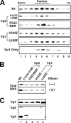
FIG. 1.
FIG. 1.
Analysis of mutant particles. (A) Particle preparations by sucrose sedimentation. Lysates prepared from cells transfected with either NO-SV40 wild type (Wt), Vp3 mutants F157E-I158E and P164R-G165E-G166R (PGG-RER), Vp1 mutants V243E and L245E, or SV-Vp1 (Vp1-Only) were treated with DNase I and sedimented through a 5 to 32% sucrose gradient. Aliquots of 10 fractions taken from the bottom of the tube were run on 10% sodium dodecyl sulfate-polyacrylamide gel electrophoresis, and Vp1 was detected by Western blotting. Vp1 peak fractions are indicated (Particles). (B and C) Viral DNA contents and capsid protein compositions of mutant particles. (B) Peak particle fractions from panel A were pooled and concentrated by pelleting them with high-speed centrifugation. Mutant particles, as well as virions, were either not treated (−) or treated (+) with 0.1 U/μl of DNase I for 20 min at 37°C; DNA was extracted, and viral DNA was detected by Southern blotting using 32P-labeled SV40 DNA as the probe. (C) Particle preparations were probed by Western blotting for the presence of either Vp1 (top) or minor capsid proteins (bottom). The positions of the bands representing the respective capsid proteins are indicated.
FIG. 2.
FIG. 2.
Analysis of Vp1-only particles. NO-SV40 (Wt) and SV-Vp1 (Vp1-Only) particles were purified and pelleted as for Fig. 1B. The Vp1 content in each preparation was examined by Western blotting (inset). The particles were incubated either in the absence (−) or in the presence (+) of EGTA and DTT (EGTA/DTT Treatment) and digested with DNase I. Viral DNA was extracted after the DNase I digestion and measured by quantitative PCR using the iCycler thermal-cycler system (Bio-Rad) and is indicated as copy numbers.
FIG. 3.
FIG. 3.
Histone contents of mutant particles. Aliquots of EGTA/DTT-disrupted particles of NO-SV40 (WT; row A), of mutant V243E (row B) or L245E (row C), or of SV-Vp1 (Vp1-only; row D) were immunoprecipitated with rabbit anti-sheep IgG (Cont), anti-histone H2B (H2B), anti-histone H3 (H3), or anti-histone H4 (H4) antibody. The respective IPs were mixed with the NO-pSV40ΔNcoI standard DNA, and their DNAs were extracted and subjected to semiquantitative PCR for viral DNA as described in Materials and Methods. A 2.2-kbp fragment (solid arrow; vDNA) and a 1.7-kbp fragment (solid arrowhead; con DNA) were generated from the amplification of the NO-SV40 genome and coextracted standard DNAs, respectively. A 1.8-kbp fragment (empty arrow) and a 2.3-kbp fragment (empty arrowhead) were generated from the amplification of Vp1-only and coextracted standard DNAs, respectively.
FIG. 4.
FIG. 4.
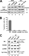
Cell entry, T-antigen expression, and protein associations of internalized viral DNA. (A) Cell attachment and internalization by mutant particles. TC-7 cells infected with particles of NO-SV40 (Wt; lanes 1 and 4), of mutant V243E (lane 2) or L245E (lane 3), or of SV-Vp1 (Vp1-Only; lane 5) were harvested either by scraping (Cell associated) or by trypsin treatment (Internalized). Viral DNA was extracted from one-eighth of the input particles (Input) or from one-half of the infected-cell lysate, linearized, and detected by Southern blotting. Similarly, the amount of Vp1 in the cells infected with either NO-SV40 (lane 4) or Vp1-only (lane 5) particles was detected by Western blotting. (B) T-antigen expression in mutant-particle-infected cells. Cells on coverslips were infected for 20 h with the same set of wild-type and mutant particles described for panel A above. The number of T-antigen-positive cells was determined by immunofluorescence microscopy. Each bar represents the average, with standard deviation, of three sets of experiments, in each of which approximately 2,000 cells were counted. (C) Analysis of virion DNA association with internalized capsid proteins and importins. Cytoplasmic fractions prepared from cells infected for 6 h with the same set of wild-type and mutant particles described for panel A were reacted with rabbit anti-mouse IgG (lane 2; Cont), anti-Vp1 (lane 3), or a mixture of anti-importin α and anti-importin β antibodies (lane 4; Imps), as described in Materials and Methods. The coimmunoprecipitated viral DNA (vDNA) was detected via semiquantitative PCR in the presence of a fixed amount of control DNA. The arrow and arrowhead designations for the amplification products are the same as those in the legend to Fig. 3. For “Input” (lane 1), one-fifth as much cytoplasmic lysate as the amount used for each immunoprecipitation was used.

References

    1. Boura, E., D. Liebl, R. Spisek, J. Fric, M. Marek, J. Stokrova, V. Holan, and J. Forstova. 2005. Polyomavirus EGFP-pseudocapsids: Analysis of model particles for introduction of proteins and peptides into mammalian cells. FEBS Lett. 579:6549-6558. - PubMed
    1. Brady, J. N., V. D. Winston, and R. A. Consigli. 1977. Dissociation of polyoma virus by the chelation of calcium ions found associated with purified virions. J. Virol. 23:717-724. - PMC - PubMed
    1. Carbone, M., A. Reale, A. Di Sauro, O. Sthandier, M. I. Garcia, R. Maione, P. Caiafa, and P. Amati. 2006. PARP-1 interaction with VP1 capsid protein regulates polyomavirus early gene expression. J. Mol. Biol. 363:773-785. - PubMed
    1. Cavaldesi, M., M. Caruso, O. Sthandier, P. Amati, and M. I. Garcia. 2004. Conformational changes of murine polyomavirus capsid proteins induced by sialic acid binding. J. Biol. Chem. 279:41573-41579. - PubMed
    1. Chang, D., C. Y. Fung, W. C. Ou, P. C. Chao, S. Y. Li, M. Wang, Y. L. Huang, T. Y. Tzeng, and R. T. Tsai. 1997. Self-assembly of the JC virus major capsid protein, VP1, expressed in insect cells. J. Gen. Virol. 78:1435-1439. - PubMed

Publication types

LinkOut - more resources