

Structures of a human papillomavirus (HPV) E6 polypeptide bound to MAGUK proteins: mechanisms of targeting tumor suppressors by a high-risk HPV oncoprotein
- PMID: 17267502
- PMCID: PMC1866053
- DOI: 10.1128/JVI.02044-06
Structures of a human papillomavirus (HPV) E6 polypeptide bound to MAGUK proteins: mechanisms of targeting tumor suppressors by a high-risk HPV oncoprotein
Abstract
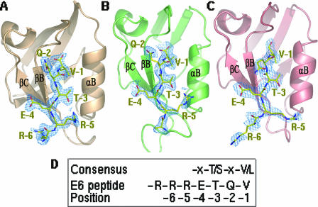
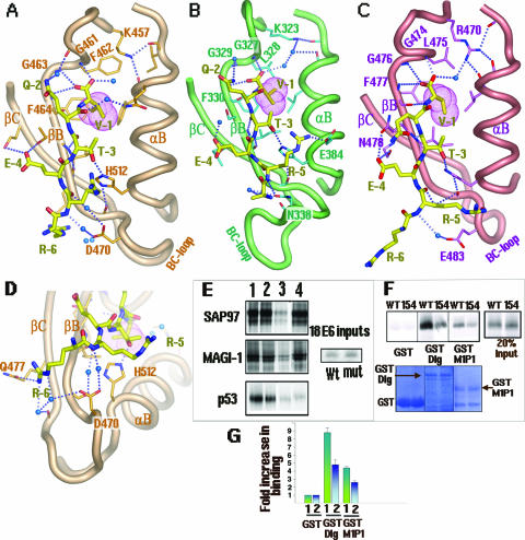
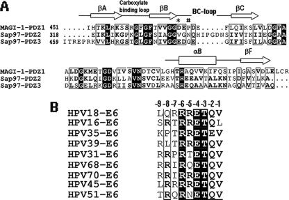
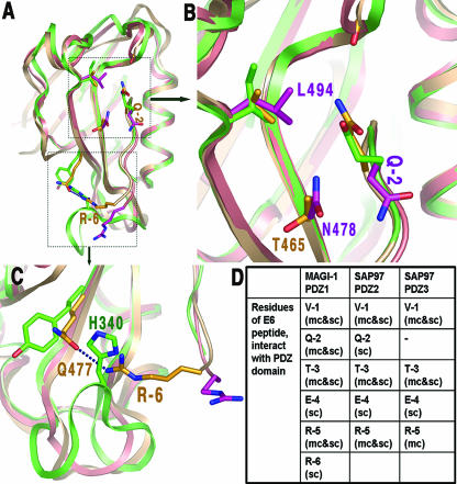
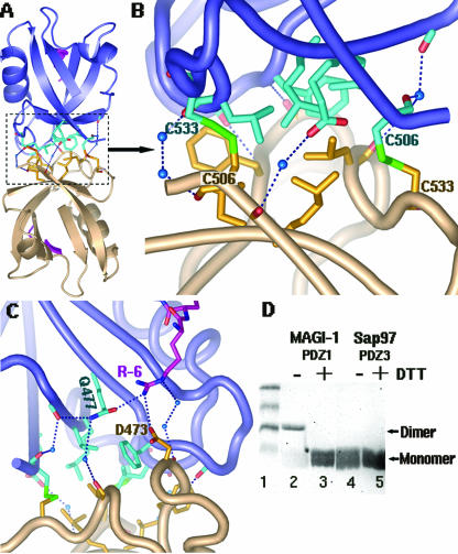
Human papillomavirus (HPV) E6 oncoprotein targets certain tumor suppressors such as MAGI-1 and SAP97/hDlg for degradation. A short peptide at the C terminus of E6 interacts specifically with the PDZ domains of these tumor suppressors, which is a property unique to high-risk HPVs that are associated with cervical cancer. The detailed recognition mechanisms between HPV E6 and PDZ proteins are unclear. To understand the specific binding of cellular PDZ substrates by HPV E6, we have solved the crystal structures of the complexes containing a peptide from HPV18 E6 bound to three PDZ domains from MAGI-1 and SAP97/Dlg. The complex crystal structures reveal novel features of PDZ peptide recognition that explain why high-risk HPV E6 can specifically target these cellular tumor suppressors for destruction. Moreover, a new peptide-binding loop on these PDZs is identified as interacting with the E6 peptide. Furthermore, we have identified an arginine residue, unique to high-risk HPV E6 but outside the canonical core PDZ recognition motif, that plays an important role in the binding of the PDZs of both MAGI-I and SAP97/Dlg, the mutation of which abolishes E6's ability to degrade the two proteins. Finally, we have identified a dimer form of MAGI-1 PDZ domain 1 in the cocrystal structure with E6 peptide, which may have functional relevance for MAGI-1 activity. In addition to its novel insights into the biochemistry of PDZ interactions, this study is important for understanding HPV-induced oncogenesis; this could provide a basis for developing antiviral and anticancer compounds.
Figures
Similar articles
-
Solution structure of the hDlg/SAP97 PDZ2 domain and its mechanism of interaction with HPV-18 papillomavirus E6 protein.Biochemistry. 2007 Sep 25;46(38):10864-74. doi: 10.1021/bi700879k. Epub 2007 Aug 22. Biochemistry. 2007. PMID: 17713926
-
Design of a PDZbody, a bivalent binder of the E6 protein from human papillomavirus.Sci Rep. 2015 Mar 23;5:9382. doi: 10.1038/srep09382. Sci Rep. 2015. PMID: 25797137 Free PMC article.
-
Chimaeric HPV E6 proteins allow dissection of the proteolytic pathways regulating different E6 cellular target proteins.Oncogene. 2002 Nov 21;21(53):8140-8. doi: 10.1038/sj.onc.1206026. Oncogene. 2002. PMID: 12444549
-
Structure and function of the papillomavirus E6 protein and its interacting proteins.Front Biosci. 2008 Jan 1;13:121-34. doi: 10.2741/2664. Front Biosci. 2008. PMID: 17981532 Review.
-
Cellular binding partners of the human papillomavirus E6 protein.Arch Virol. 2008;153(3):397-408. doi: 10.1007/s00705-007-0022-5. Epub 2008 Jan 3. Arch Virol. 2008. PMID: 18172569 Free PMC article. Review.
Cited by
-
Novel phospho-switch function of delta-catenin in dendrite development.J Cell Biol. 2020 Nov 2;219(11):e201909166. doi: 10.1083/jcb.201909166. J Cell Biol. 2020. PMID: 33007084 Free PMC article.
-
SAP97 controls the trafficking and resensitization of the beta-1-adrenergic receptor through its PDZ2 and I3 domains.PLoS One. 2013 May 16;8(5):e63379. doi: 10.1371/journal.pone.0063379. Print 2013. PLoS One. 2013. PMID: 23696820 Free PMC article.
-
Papillomavirus E6 oncoproteins.Virology. 2013 Oct;445(1-2):115-37. doi: 10.1016/j.virol.2013.04.026. Epub 2013 May 24. Virology. 2013. PMID: 23711382 Free PMC article. Review.
-
Analysis of the PDZ binding specificities of Influenza A virus NS1 proteins.Virol J. 2011 Jan 19;8:25. doi: 10.1186/1743-422X-8-25. Virol J. 2011. PMID: 21247458 Free PMC article.
-
Pharmacophore Modeling and Molecular Docking Studies of potential inhibitors to E6 PBM-PDZ from Human Papilloma Virus (HPV).Bioinformation. 2015 Aug 31;11(8):401-6. doi: 10.6026/97320630011401. eCollection 2015. Bioinformation. 2015. PMID: 26420921 Free PMC article.
References
-
- Reference deleted.
-
- Berger, T., E. Stockfleth, T. Meyer, F. Kiesewetter, and J. O. Funk. 2005. Multiple disseminated large-cell acanthomas of the skin associated with human papillomavirus type 6. J. Am. Acad. Dermatol. 53:335-337. - PubMed
-
- Brenman, J. E., D. S. Chao, S. H. Gee, A. W. McGee, S. E. Craven, D. R. Santillano, Z. Wu, F. Huang, H. Xia, M. F. Peters, S. C. Froehner, and D. S. Bredt. 1996. Interaction of nitric oxide synthase with the postsynaptic density protein PSD-95 and alpha1-syntrophin mediated by PDZ domains. Cell 84:757-767. - PubMed
Publication types
MeSH terms
Substances
Grants and funding
LinkOut - more resources
Full Text Sources
Other Literature Sources
Molecular Biology Databases
