Mutation in the glycosylated gag protein of murine leukemia virus results in reduced in vivo infectivity and a novel defect in viral budding or release
- PMID: 17267509
- PMCID: PMC1866097
- DOI: 10.1128/JVI.01538-06
Mutation in the glycosylated gag protein of murine leukemia virus results in reduced in vivo infectivity and a novel defect in viral budding or release
Abstract
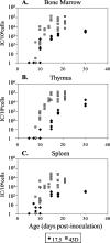
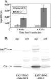
All gammaretroviruses, including murine leukemia viruses (MuLVs), feline leukemia viruses, and gibbon-ape leukemia virus, encode an alternate, glycosylated form of Gag polyprotein (glyco-Gag or gPr80gag) in addition to the polyprotein precursor of the viral capsid proteins (Pr65gag). gPr80gag is translated from an upstream in-frame CUG initiation codon, in contrast to the AUG codon used for Pr65gag. The role of glyco-Gag in MuLV replication has been unclear, since gPr80gag-negative Moloney MuLV (M-MuLV) mutants are replication competent in vitro and pathogenic in vivo. However, reversion to the wild type is frequently observed in vivo. In these experiments, in vivo inoculation of a gPr80gag mutant, Ab-X-M-MuLV, showed substantially lower (2 log) initial infectivity in newborn NIH Swiss mice than that of wild-type virus, and revertants to the wild type could be detected by PCR cloning and DNA sequencing as early as 15 days postinfection. Atomic force microscopy of Ab-X-M-MuLV-infected producer cells or of the PA317 amphotropic MuLV-based vector packaging line (also gPr80gag negative) revealed the presence of tube-like viral structures on the cell surface. In contrast, wild-type virus-infected cells showed the typical spherical, 145-nm particles observed previously. Expression of gPr80gag in PA317 cells converted the tube-like structures to typical spherical particles. PA317 cells expressing gPr80gag produced 5- to 10-fold more infectious vector or viral particles as well. Metabolic labeling studies indicated that this reflected enhanced virus particle release rather than increased viral protein synthesis. These results indicate that gPr80gag is important for M-MuLV replication in vivo and in vitro and that the protein may be involved in a late step in viral budding or release.
Figures


Similar articles
-
Moloney murine leukemia virus glyco-gag facilitates xenotropic murine leukemia virus-related virus replication through human APOBEC3-independent mechanisms.Retrovirology. 2012 Jul 24;9:58. doi: 10.1186/1742-4690-9-58. Retrovirology. 2012. PMID: 22828015 Free PMC article.
-
Construction and characterization of Moloney murine leukemia virus mutants unable to synthesize glycosylated gag polyprotein.Proc Natl Acad Sci U S A. 1983 Oct;80(19):5965-9. doi: 10.1073/pnas.80.19.5965. Proc Natl Acad Sci U S A. 1983. PMID: 6310608 Free PMC article.
-
Murine leukemia virus glycosylated Gag (gPr80gag) facilitates interferon-sensitive virus release through lipid rafts.Proc Natl Acad Sci U S A. 2010 Jan 19;107(3):1190-5. doi: 10.1073/pnas.0908660107. Epub 2009 Dec 28. Proc Natl Acad Sci U S A. 2010. PMID: 20080538 Free PMC article.
-
Mutants of murine leukemia viruses and retroviral replication.Biochim Biophys Acta. 1987 Jul 8;907(2):93-123. doi: 10.1016/0304-419x(87)90001-1. Biochim Biophys Acta. 1987. PMID: 3036230 Review.
-
HTLV-1 structural proteins.Virus Res. 2001 Oct 30;78(1-2):5-16. doi: 10.1016/s0168-1702(01)00278-7. Virus Res. 2001. PMID: 11520576 Review.
Cited by
-
Exploring FeLV-Gag-Based VLPs as a New Vaccine Platform-Analysis of Production and Immunogenicity.Int J Mol Sci. 2023 May 19;24(10):9025. doi: 10.3390/ijms24109025. Int J Mol Sci. 2023. PMID: 37240371 Free PMC article.
-
Human and murine APOBEC3s restrict replication of koala retrovirus by different mechanisms.Retrovirology. 2015 Aug 8;12:68. doi: 10.1186/s12977-015-0193-1. Retrovirology. 2015. PMID: 26253512 Free PMC article.
-
Deaminase-Dead Mouse APOBEC3 Is an In Vivo Retroviral Restriction Factor.J Virol. 2018 May 14;92(11):e00168-18. doi: 10.1128/JVI.00168-18. Print 2018 Jun 1. J Virol. 2018. PMID: 29593034 Free PMC article.
-
N-linked glycosylation protects gammaretroviruses against deamination by APOBEC3 proteins.J Virol. 2015 Feb;89(4):2342-57. doi: 10.1128/JVI.03330-14. Epub 2014 Dec 10. J Virol. 2015. PMID: 25505062 Free PMC article.
-
Murine leukemia virus glycosylated Gag blocks apolipoprotein B editing complex 3 and cytosolic sensor access to the reverse transcription complex.Proc Natl Acad Sci U S A. 2013 May 28;110(22):9078-83. doi: 10.1073/pnas.1217399110. Epub 2013 May 13. Proc Natl Acad Sci U S A. 2013. PMID: 23671100 Free PMC article.
References
-
- Chun, R., and H. Fan. 1994. Recovery of glycosylated gag virus from mice infected with a glycosylated gag-negative mutant of Moloney murine leukemia virus. J. Biomed. Sci. 1:218-223. - PubMed
Publication types
MeSH terms
Substances
Grants and funding
LinkOut - more resources
Full Text Sources
Research Materials
Miscellaneous

