Comparative Study
. 2007 Jan 31;27(5):1033-44.
doi: 10.1523/JNEUROSCI.3160-06.2007.

Postsynaptic membrane addition depends on the Discs-Large-interacting t-SNARE Gtaxin

Affiliations
Comparative Study

Postsynaptic membrane addition depends on the Discs-Large-interacting t-SNARE Gtaxin

David Gorczyca et al. J Neurosci. .

Abstract

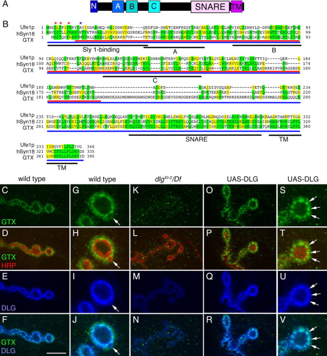
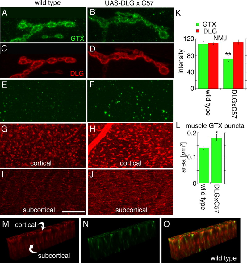
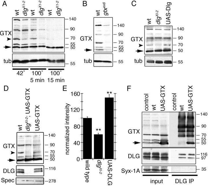
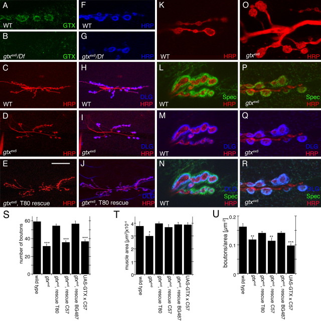
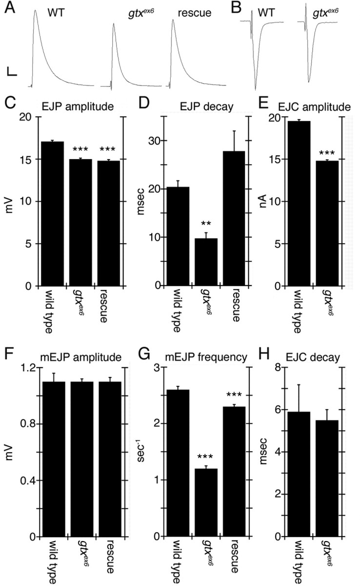
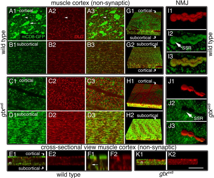
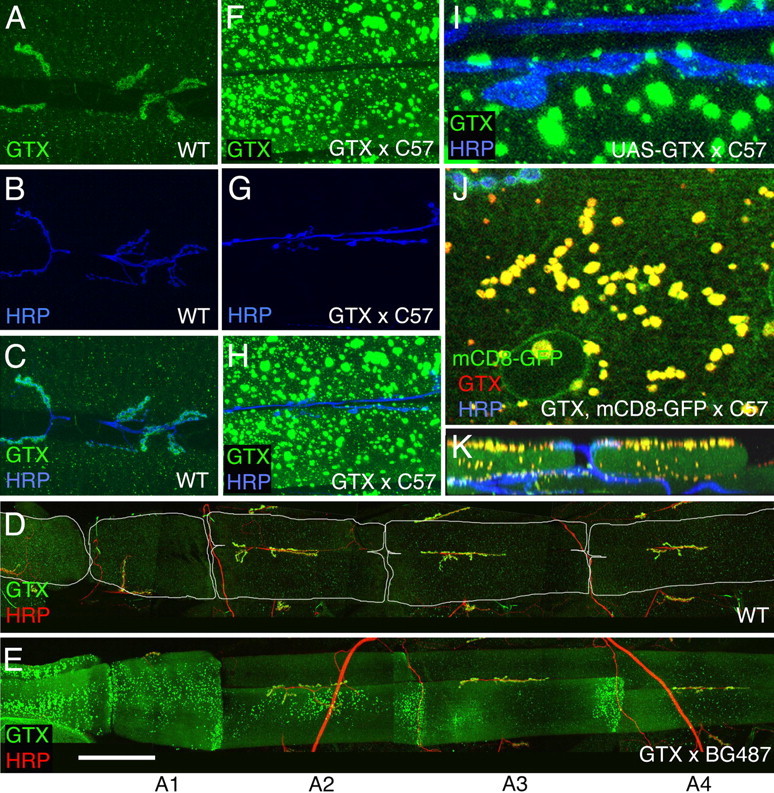
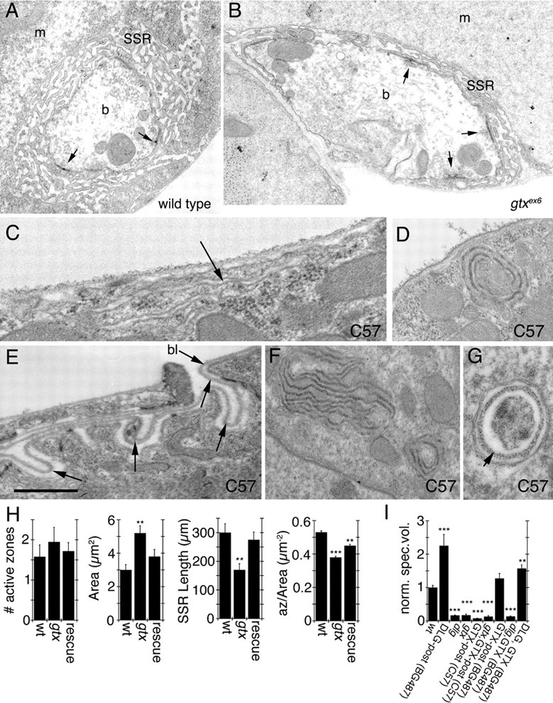
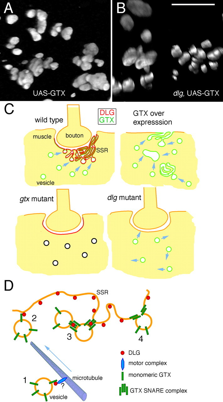
Targeted membrane addition is a hallmark of many cellular functions. In the nervous system, modification of synaptic membrane size has a major impact on synaptic function. However, because of the complex shape of neurons and the need to target membrane addition to very small and polarized synaptic compartments, this process is poorly understood. Here, we show that Gtaxin (GTX), a Drosophila t-SNARE (target-soluble N-ethylmaleimide-sensitive factor attachment protein receptor), is required for expansion of postsynaptic membranes during new synapse formation. Mutations in gtx lead to drastic reductions in postsynaptic membrane surface, whereas gtx upregulation results in the formation of complex membrane structures at ectopic sites. Postsynaptic GTX activity depends on its direct interaction with Discs-Large (DLG), a multidomain scaffolding protein of the PSD-95 (postsynaptic density protein-95) family with key roles in cell polarity and formation of cellular junctions as well as synaptic protein anchoring and trafficking. We show that DLG selectively determines the postsynaptic distribution of GTX to type I, but not to type II or type III boutons on the same cell, thereby defining sites of membrane addition to this unique set of glutamatergic synapses. We provide a mechanistic explanation for selective targeted membrane expansion at specific synaptic junctions.


Figures

Figure 1.
Figure 1.
GTX protein structure and synaptic localization. A, Organization of GTX into protein domains. (N, N-terminal homologous Sly1 binding site; A, B, C, predicted Ha, Hb, and Hc domains; SNARE, coiled-coil SNARE domain; TM, transmembrane domain). B, Protein alignment of yeast Ufe1p, human Syntaxin 18, and GTX. Asterisks represent amino acids required for Sly1 binding (red, conserved; blue, not conserved). Regions of identity are highlighted in yellow, whereas conserved regions are marked in green. Black underlining indicates different protein domains. Red and blue underlining shows the GTX protein regions used to generate the anti-GTX antisera. C–V, Single confocal slices of third instar larval NMJs labeled with anti-GTX (green), anti-HRP (red), and anti-DLG (blue), shown at low (C–F, K–R) and high (G–J, S–V) magnification in wild type (C–J), dlg XI2/Df(dlg) (K–N), and UAS-DLG (O–V) overexpressed using the BG487 Gal4. The arrow in G–J points to a budding bouton. Arrows in S–V point to accumulations of DLG, which are accompanied by accumulations of GTX. Scale bar: C–F, K–N, O–R, 7 μm; G–J, S–V, 3 μm.
Figure 2.
Figure 2.
Effects of strong overexpression of DLG using the Gal4 driver C57. A–D, Single confocal slices of third instar larval NMJs stained with antibodies against GTX (green) and DLG (red) in wild type (A, C) and larvae overexpressing DLG (B, D) with the C57 Gal4 driver. E–J, Single confocal slices of an extrasynaptic region of the larval muscles showing the localization of GTX (green) and DLG (red) at the cortical region (E–H) and the subcortical region (I, J) of a muscle cell. K, L, Graphs showing the intensity of the DLG and GTX label at the synapse (K) and the area of GTX-labeled puncta in muscles (L) of wild-type and larvae overexpressing DLG using the C57 Gal4 driver (n = 12 for each genotype). M–O, GTX and DLG immunoreactivity in the regions between the cortical and subcortical DLG networks seen in G–J. *p < 0.05; **p < 0.001. Scale bar: A–D, 5 μm; E–J, 8 μm; M–O, 17 μm.
Figure 3.
Figure 3.
GTX forms SDS-resistant high molecular weight complexes, and DLG regulates these complexes and is associated with GTX in the body wall muscles. A, Western blot of wild-type and dlg XI−2 mutant body wall muscle extracts subjected to different temperatures to demonstrate the presence of SDS-resistant high molecular weight complexes and the regulation of some of these complexes by DLG. B, Western blot of wild-type and gtx ex6 mutant extracts incubated at 42°C in loading buffer. C, Western blot of body wall muscle extracts from wild type, dlg XI2, and larvae overexpressing DLG incubated at 42°C in loading buffer. D, Western blot of wild type, overexpression of GTX in a dlg XI2 mutant background, and overexpression of GTX in a wild-type background. E, Intensity of the GTX 70 kDa band (A, C, D, arrowhead) observed in dlg mutants and after overexpressing DLG, normalized to wild-type intensity. n = 3 independent Western blots. F, Immunoprecipitation of body wall muscle extracts using anti-DLG antibodies. The blots were sequentially probed with antibodies to GTX, DLG, and Syntaxin-1A. Inputs represent 10% of the extract used for immunoprecipitations. Numbers at the right of the blots represent molecular weights in kDa. The arrow in A–D and F indicates GTX monomer. The arrowhead in A, C, and D indicates the 70 kDa GTX complex. tub, Tubulin; Spec, Spectrin; IP, immunoprecipitation.
Figure 4.
Figure 4.
Abnormal NMJs in gtx mutants. A–R, Confocal images of third instar larval NMJs stained with anti-GTX (green) and anti-HRP (blue) (A, B, F, G), anti-HRP (red) and anti-DLG (blue) (C–E, H–J), anti-HRP (red) (K, O) and anti-Spectrin (Spec) (green), anti-DLG (blue), and anti-HRP (red) (L–N, P–R) in wild-type (WT) (A, C, F, H, K–N), gtx ex6 (B, D, G, I, O–R), and gtx ex6 (E, J) mutants expressing transgenic GTX using the T80 Gal4 driver. S–U, Morphometric analysis at muscles 6 and 7 in the genotypes indicated, showing number of synaptic boutons (S), muscle surface area (T), and number of boutons normalized by the muscle area (U) (n = 12 for each genotype). *p < 0.05; **p < 0.001; ***p < 0.0001. Scale bar: A, B, F, G, K–R, 7 μm; C–E, H–J, 35 μm.
Figure 5.
Figure 5.
Electrophysiological defects in gtx mutants. A, Representative EJP traces of wild-type, gtx ex6, and gtx transgene expression in a gtx ex6 mutant background using the T80 Gal4 driver. B, Representative voltage-clamp EJC recordings of wild-type and gtx ex6 mutants. C–H, quantification of EJP amplitude (C), EJP decay time constant (D), EJC amplitude (E), mEJP amplitude (F), mEJP frequency (G), and EJC decay constant (H). n = 7 for all genotypes. **p < 0.001; ***p < 0.0001. Calibration: A, 2 mV, 40 ms; B, 3 nA, 15 ms.
Figure 6.
Figure 6.
Muscle endomembrane system in wild-type and gtx ex6 larvae. Views of muscle extrasynaptic regions (A1–D3, G1–H2) and of synaptic boutons (I1–J3) in preparations labeled with mCD8-GFP (green) and anti-DLG (red) in wild type (A1–B3, E1–G2, I1–I3) and gtxex6 (C1–D3, H1, H2, J1–K2). A1, Single confocal slices at the muscle cortex, near the muscle surface (A3, C1–C3), and single confocal slices at the subcortical muscle region, below the nuclei (B1–B3, D1–D3). G1, G2, H1, H2, Three-dimensional rendering of confocal slices spanning the muscle cortex and part of the subcortical region shown from the surface of the muscle (G1, H1) and from the subcortical muscle region (G2, H2). E1–F2, K1, K2, Transversal section through a confocal stack showing the relationship between the cortical and subcortical networks. n, Nucleus. F1, F2, A high-magnification view of the muscle area between the cortical and subcortical networks, showing structures connecting the two networks. I1–I3, J1–J3, Three-dimensional rendering of an NMJ branch. mCD8-GFP labels muscle membranes including the SSR, and anti-DLG antibodies label mostly the SSR. Cortical, Below plasma membrane; subcortical, below nuclei. Scale bar: A1–D3, 12 μm; E1, E2, G1–H2, K1, K2, 20 μm; I1–I3, J1–J3, 10 μm; F1, F2, 4 μm.
Figure 7.
Figure 7.
Effects of overexpressing GTX in larval muscles. A–K, Confocal images of larval NMJs in wild type (A–D) and larvae overexpressing transgenic GTX (E–K) in muscles using the Gal4 driver BG487 (D, E) (weak muscle expression in A3) and C57 (F–K) (strong pan-muscle expression), stained with antibodies to HRP (blue) and GTX (green) (A–C, F–I), GTX (green) and HRP (red) (D, E), and GTX (red), HRP (blue), and mCD8-GFP (green) (J, K). A–C and F–K are images from muscles 6 and 7 in A3, whereas D and E show abdominal segments A1–A4. K is a transversal section through a confocal stack showing the localization of GTX-containing large vesicles at the muscle apical region. Scale bar: A–C, F–H, 50 μm; D, E, 185 μm; I, 8 μm; J, 12 μm; K, 30 μm.
Figure 8.
Figure 8.
Ultrastructure of synaptic boutons and muscles in gtx mutants. A–G, Electron micrographs of type Ib synaptic boutons (A, B) in wild type (A) and gtx ex6 mutants (B). m, Muscle; b, bouton. Arrows point to T-bar active zones. C–G, Extrasynaptic membrane structures observed in larvae overexpressing GTX using the strong pan-muscle Gal4 driver C57. Arrows in C and E point to surface membranous structures resembling the SSR, and the arrow in G points to electron dense material resembling basal lamina (bl). H, Morphometric analysis of synaptic boutons in wild type, gtx ex6 mutants, and gtx ex6 mutants expressing transgenic GTX using the Gal4 strain BG487. Numbers analyzed were 12 boutons in two wild-type larvae, 19 boutons in two gtx ex6, and 12 boutons in three gtx ex6 expressing GTX in muscles using the BG487 Gal4. I, Quantification of the volume occupied by Spectrin at the postsynaptic area in different genotypes. n = 6 samples for each phenotype; **p = 0.001; ***p < 0.0001. n = 6 for gtx ex6 and gtx ex6; C57 and 8 for all other genotypes. Scale bar: A, B, 1.5 μm; C, 0.2 μm; D, F, 0.5 μm; E, G, 0.3.
Figure 9.
Figure 9.
Ectopic SSR formation after strong GTX overexpression and proposed function of DLG and GTX during targeted membrane addition. A, B, Three-dimensional renderings of ectopic GTX-positive structures observed after overexpressing GTX with the strong C57 driver in wild type (A) and dlg XI2 mutant (B) backgrounds. Scale bar: A, B, 8 μm. C, Model of membrane trafficking to the postsynaptic region. In wild type, the presence of DLG at the postsynaptic membrane directs the fusion of GTX-positive vesicles to this site, forming the SSR. In dlg mutants, GTX targeting to the postsynaptic membrane is perturbed, resulting in reduced SSR expansion. When GTX is severely reduced in gtx mutants, despite the relatively normal synaptic DLG localization, fusion events are inhibited, and the SSR does not expand. When GTX is expressed at very high levels, inappropriate homotypic fusion occurs, leading to the formation of large vesicles and SSR-like structures in the cytoplasm and extrasynaptic muscle surface. D, Potential interactions between DLG and GTX during SSR formation. 1, GTX-positive vesicles are trafficked to the SSR, perhaps in a DLG dependent manner. 2, Through low-affinity interactions between DLG and GTX monomers, GTX-containing vesicles are concentrated at the SSR region. 3, Increased concentration of GTX-containing vesicles facilitates the formation of SNARE complexes, which are stabilized/protected by DLG. 4, The GTX SNARE complex formations allow the fusion of GTX vesicles with the SSR membrane, thus resulting in SSR expansion.

Similar articles

Cited by

References

    1. Ashley J, Packard M, Ataman B, Budnik V. Fasciclin II signals new synapse formation through amyloid precursor protein and the scaffolding protein dX11/Mint. J Neurosci. 2005;25:5943–5955. - PMC - PubMed
    1. Budnik V, Zhong Y, Wu CF. Morphological plasticity of motor axons in Drosophila mutants with altered excitability. J Neurosci. 1990;10:3754–3768. - PMC - PubMed
    1. Budnik V, Koh YH, Guan B, Hartmann B, Hough C, Woods D, Gorczyca M. Regulation of synapse structure and function by the Drosophila tumor suppressor gene dlg. Neuron. 1996;17:627–640. - PMC - PubMed
    1. Cathala L, Holderith NB, Nusser Z, DiGregorio DA, Cull-Candy SG. Changes in synaptic structure underlie the developmental speeding of AMPA receptor-mediated EPSCs. Nat Neurosci. 2005;8:1310–1318. - PubMed
    1. El-Husseini AE, Schnell E, Chetkovich DM, Nicoll RA, Bredt DS. PSD-95 involvement in maturation of excitatory synapses. Science. 2000a;290:1364–1368. - PubMed

Publication types

MeSH terms

Associated data

LinkOut - more resources