

Neurophysiological mechanisms involved in transfer of procedural knowledge
- PMID: 17267558
- PMCID: PMC6673204
- DOI: 10.1523/JNEUROSCI.4128-06.2007
Neurophysiological mechanisms involved in transfer of procedural knowledge
Abstract
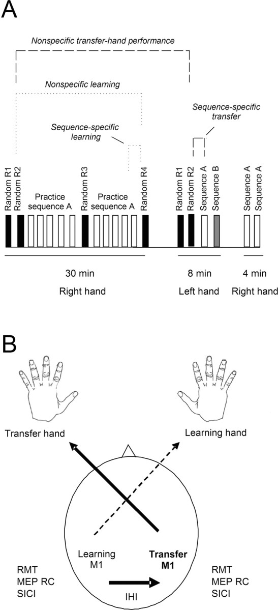
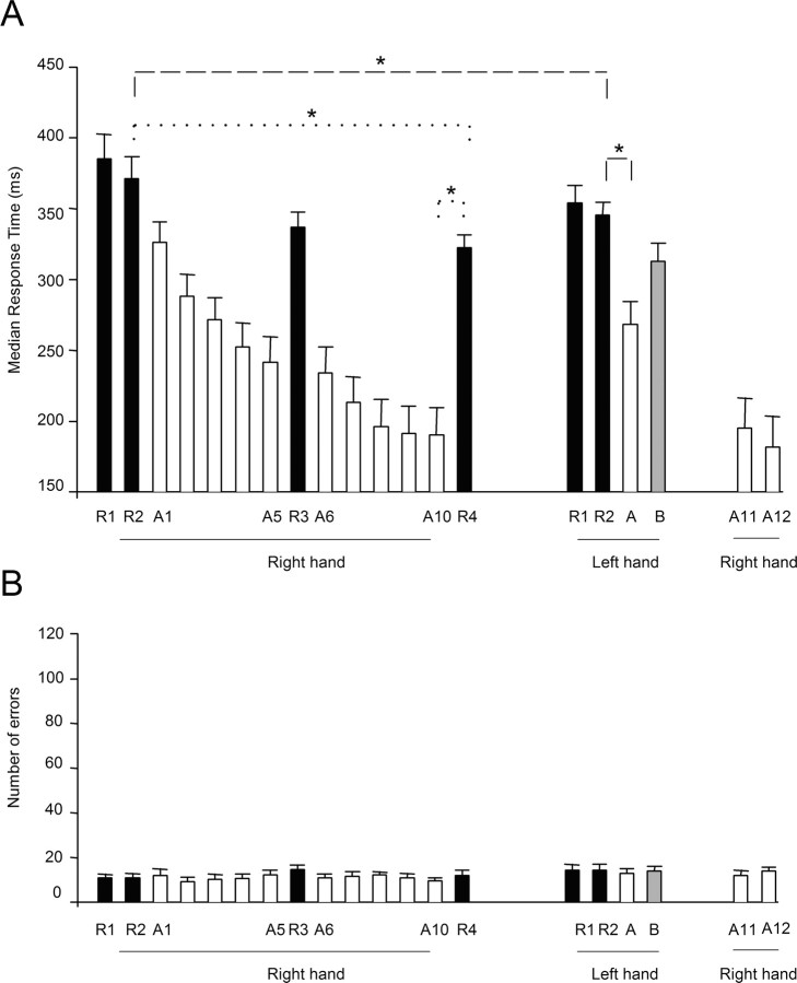
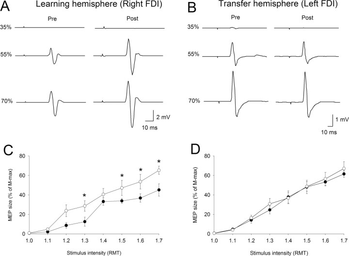
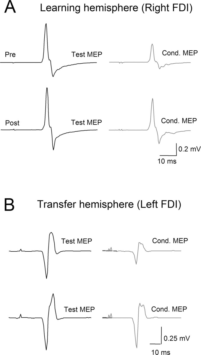
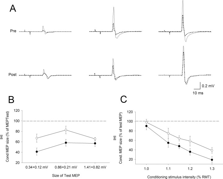
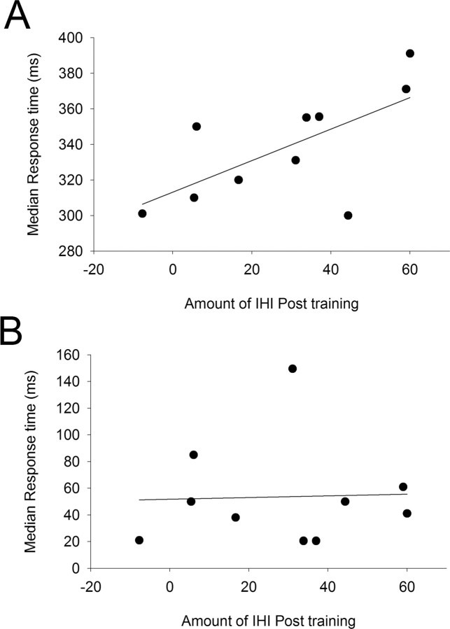
Learning to perform a motor task with one hand results in performance improvements in the other hand, a process called intermanual transfer. To gain information on its neural mechanisms, we studied this phenomenon using the serial reaction-time task (SRTT). Sixteen, right-handed volunteers trained a 12-item sequence of key presses repeated without the subjects' knowledge. Blocks with no repeating sequence, called random blocks, were interspersed with sequence-training blocks. Response times improved in random and training blocks in both hands. The former result reflects nonspecific improvement in performance, and the latter represents a sequence-specific improvement. To evaluate changes in the primary motor cortex (M1), we tested resting motor thresholds (RMT), recruitments curves to transcranial magnetic stimulation (RC), short intracortical inhibition (SICI), and interhemispheric inhibition (IHI) from the dominant left (learning) to the nondominant right (transfer) hemisphere, before and after SRTT training. Training resulted in (1) increased RC and decreased SICI but no changes in RMT in the learning hemisphere, (2) decreased SICI and no changes in RC or RMT in the transfer hemisphere, and (3) decreased IHI. The amount in IHI after training correlated with nonspecific performance improvements in the transfer hand but not with sequence-specific performance improvements. Our results indicate that modulation of interhemispheric inhibition between the M1 areas may, as a result of the learning that has occurred in one hemisphere after practice with one hand, contribute to faster, more skilled performance of the opposite hand.
Figures
Similar articles
-
Mechanisms controlling motor output to a transfer hand after learning a sequential pinch force skill with the opposite hand.Clin Neurophysiol. 2009 Oct;120(10):1859-65. doi: 10.1016/j.clinph.2009.08.013. Epub 2009 Sep 18. Clin Neurophysiol. 2009. PMID: 19766535 Free PMC article.
-
Mechanisms underlying functional changes in the primary motor cortex ipsilateral to an active hand.J Neurosci. 2008 May 28;28(22):5631-40. doi: 10.1523/JNEUROSCI.0093-08.2008. J Neurosci. 2008. PMID: 18509024 Free PMC article.
-
Intermanual Differences in movement-related interhemispheric inhibition.J Cogn Neurosci. 2007 Feb;19(2):204-13. doi: 10.1162/jocn.2007.19.2.204. J Cogn Neurosci. 2007. PMID: 17280510
-
Motor learning and cross-limb transfer rely upon distinct neural adaptation processes.J Neurophysiol. 2016 Aug 1;116(2):575-86. doi: 10.1152/jn.00225.2016. Epub 2016 May 11. J Neurophysiol. 2016. PMID: 27169508 Free PMC article.
-
BDNF Val66Met polymorphism is associated with altered activity-dependent modulation of short-interval intracortical inhibition in bilateral M1.PLoS One. 2018 Jun 1;13(6):e0197505. doi: 10.1371/journal.pone.0197505. eCollection 2018. PLoS One. 2018. PMID: 29856758 Free PMC article.
Cited by
-
Functional consequences of a section of the anterior part of the body of the corpus callosum: evidence from an interhemispheric transcallosal approach.J Neurol. 2012 Sep;259(9):1860-7. doi: 10.1007/s00415-012-6421-x. J Neurol. 2012. PMID: 22289969
-
High-frequency neuromuscular electrical stimulation modulates interhemispheric inhibition in healthy humans.J Neurophysiol. 2017 Jan 1;117(1):467-475. doi: 10.1152/jn.00355.2016. Epub 2016 Nov 9. J Neurophysiol. 2017. PMID: 27832594 Free PMC article.
-
A robot-aided visuomotor wrist training induces gains in proprioceptive and movement accuracy in the contralateral wrist.Sci Rep. 2021 Mar 5;11(1):5281. doi: 10.1038/s41598-021-84767-9. Sci Rep. 2021. PMID: 33674684 Free PMC article.
-
Direct and crossed effects of somatosensory electrical stimulation on motor learning and neuronal plasticity in humans.Eur J Appl Physiol. 2015 Dec;115(12):2505-19. doi: 10.1007/s00421-015-3248-z. Epub 2015 Sep 3. Eur J Appl Physiol. 2015. PMID: 26335625 Free PMC article.
-
Learning a covert sequence of effector movements: limits to its acquisition.Psychol Res. 2024 Feb;88(1):197-206. doi: 10.1007/s00426-023-01855-3. Epub 2023 Jul 9. Psychol Res. 2024. PMID: 37422801 Free PMC article.
References
-
- Almeida R, Stetter M. Modeling the link between functional imaging and neuronal activity: synaptic metabolic demand and spike rates. NeuroImage. 2002;17:1065–1079. - PubMed
-
- Andres FG, Mima T, Schulman AE, Dichgans J, Hallett M, Gerloff C. Functional coupling of human cortical sensorimotor areas during bimanual skill acquisition. Brain. 1999;122:855–870. - PubMed
-
- Ashe J, Lungu OV, Basford AT, Lu X. Cortical control of motor sequences. Curr Opin Neurobiol. 2006;16:213–221. - PubMed
-
- Bapi RS, Doya K, Harner AM. Evidence for effector independent and dependent representations and their differential time course of acquisition during motor sequence learning. Exp Brain Res. 2000;132:149–162. - PubMed
-
- Ben-Shaul Y, Drori R, Asher I, Stark E, Nadasdy Z, Abeles M. Neuronal activity in motor cortical areas reflects the sequential context of movement. J Neurophysiol. 2004;91:1748–1762. - PubMed
Publication types
MeSH terms
Grants and funding
LinkOut - more resources
Full Text Sources