

Impaired axonal regeneration by isolectin B4-binding dorsal root ganglion neurons in vitro
- PMID: 17267575
- PMCID: PMC6673184
- DOI: 10.1523/JNEUROSCI.5089-06.2007
Impaired axonal regeneration by isolectin B4-binding dorsal root ganglion neurons in vitro
Abstract
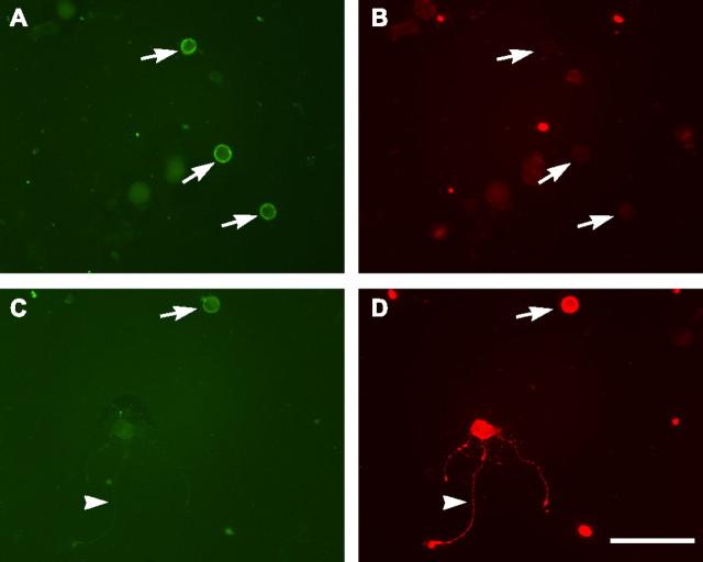
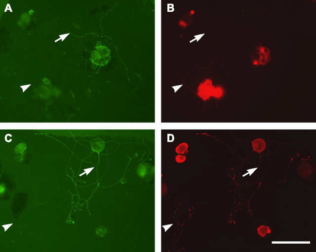
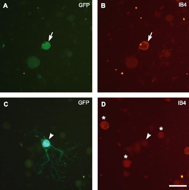
The subpopulation of dorsal root ganglion (DRG) neurons recognized by Griffonia simplicifolia isolectin B4 (IB4) differ from other neurons by expressing receptors for glial cell line-derived neurotrophic factor (GDNF) rather than neurotrophins. Additionally, IB4-labeled neurons do not express the laminin receptor, alpha7-integrin (Gardiner et al., 2005), necessary for optimal axonal regeneration in the peripheral nervous system. In cultures of dissociated DRG neurons of adult mice on laminin, robust spontaneous neurite outgrowth from IB4-negative neurons occurs and is strongly enhanced by previous axotomy. In contrast, IB4-labeled neurons show little neurite outgrowth and do not express GAP 43, even after axotomy or culture with GDNF. Moreover, growth of their axons through collagen gels is impaired compared with other DRG neurons. To determine whether the sparse neurite outgrowth of IB4-labeled neurons is attributable to lack of integrin expression, DRG cultures were infected with a herpes simplex 1 vector encoding alpha7-integrin, but its forced expression failed to promote neurite outgrowth in either IB4-labeled or other DRG neurons or in cultured adult retinal ganglion cells. Forced coexpression of both alpha7-integrin and GAP 43 also failed to promote neurite outgrowth in IB4-labeled neurons. In addition, cultured sciatic nerve segments were found to release much lower levels of GDNF, demonstrated by ELISA, than nerve growth factor. These findings together with their impaired intrinsic axonal regeneration capacity may contribute to the known vulnerability of the IB4-labeled population of DRG neurons to peripheral nerve injury.
Figures


Similar articles
-
Pericellular Griffonia simplicifolia I isolectin B4-binding ring structures in the dorsal root ganglia following peripheral nerve injury in rats.J Comp Neurol. 2001 Oct 22;439(3):259-74. doi: 10.1002/cne.1349. J Comp Neurol. 2001. PMID: 11596053
-
GDNF promotes neurite outgrowth and upregulates galectin-1 through the RET/PI3K signaling in cultured adult rat dorsal root ganglion neurons.Neurochem Int. 2013 Feb;62(3):330-9. doi: 10.1016/j.neuint.2013.01.008. Epub 2013 Jan 20. Neurochem Int. 2013. PMID: 23340048
-
Identification of some lectin IB4 binding proteins in rat dorsal root ganglia.Neuroreport. 2004 Aug 6;15(11):1705-9. doi: 10.1097/01.wnr.0000136037.54095.64. Neuroreport. 2004. PMID: 15257131
-
Laminin and growth factor receptor activation stimulates differential growth responses in subpopulations of adult DRG neurons.Eur J Neurosci. 2006 Aug;24(3):676-90. doi: 10.1111/j.1460-9568.2006.04963.x. Eur J Neurosci. 2006. PMID: 16930399
-
KV4 channels in isolectin B4 muscle dorsal root ganglion neurons of rats with experimental peripheral artery disease: effects of bradykinin B1 and B2 receptors.Am J Physiol Regul Integr Comp Physiol. 2022 Nov 1;323(5):R616-R627. doi: 10.1152/ajpregu.00117.2022. Epub 2022 Sep 12. Am J Physiol Regul Integr Comp Physiol. 2022. PMID: 36094447 Free PMC article.
Cited by
-
PTEN inhibition to facilitate intrinsic regenerative outgrowth of adult peripheral axons.J Neurosci. 2010 Jul 7;30(27):9306-15. doi: 10.1523/JNEUROSCI.6271-09.2010. J Neurosci. 2010. PMID: 20610765 Free PMC article.
-
Simultaneous Knockdown of Sprouty2 and PTEN Promotes Axon Elongation of Adult Sensory Neurons.Front Cell Neurosci. 2020 Jan 21;13:583. doi: 10.3389/fncel.2019.00583. eCollection 2019. Front Cell Neurosci. 2020. PMID: 32038175 Free PMC article.
-
An in vitro assay to study induction of the regenerative state in sensory neurons.Exp Neurol. 2015 Jan;263:350-63. doi: 10.1016/j.expneurol.2014.10.012. Epub 2014 Nov 4. Exp Neurol. 2015. PMID: 25447942 Free PMC article.
-
Regeneration of diabetic axons is enhanced by selective knockdown of the PTEN gene.Brain. 2014 Apr;137(Pt 4):1051-67. doi: 10.1093/brain/awu031. Epub 2014 Feb 27. Brain. 2014. PMID: 24578546 Free PMC article.
-
Proregenerative properties of ECM molecules.Biomed Res Int. 2013;2013:981695. doi: 10.1155/2013/981695. Epub 2013 Sep 9. Biomed Res Int. 2013. PMID: 24195084 Free PMC article. Review.
References
-
- Bailey AL, Ribeiro-da-Silva A. Transient loss of terminals from non-peptidergic nociceptive fibers in the substantia gelatinosa of spinal cord following chronic constriction injury of the sciatic nerve. Neuroscience. 2006;138:675–690. - PubMed
-
- Baloh RH, Enomoto H, Johnson EM, Jr, Milbrandt J. The GDNF family ligands and receptors: implications for neural development. Curr Opin Neurobiol. 2000;10:103–110. - PubMed
-
- Bao L, Wang HF, Cai HJ, Tong YG, Jin SX, Lu YJ, Grant G, Hokfelt T, Zhang X. Peripheral axotomy induces only very limited sprouting of coarse myelinated afferents into inner lamina II of rat spinal cord. Eur J Neurosci. 2002;16:175–185. - PubMed
-
- Bar KJ, Saldanha GJ, Kennedy AJ, Facer P, Birch R, Carlstedt T, Anand P. GDNF and its receptor component Ret in injured human nerves and dorsal root ganglia. NeuroReport. 1998;9:43–47. - PubMed
Publication types
MeSH terms
Substances
Grants and funding
LinkOut - more resources
Full Text Sources
Other Literature Sources
Miscellaneous