Comparative Study
. 2007 Jan 31;27(5):1211-9.
doi: 10.1523/JNEUROSCI.4159-06.2007.

Presynaptic monoacylglycerol lipase activity determines basal endocannabinoid tone and terminates retrograde endocannabinoid signaling in the hippocampus

Affiliations
Comparative Study

Presynaptic monoacylglycerol lipase activity determines basal endocannabinoid tone and terminates retrograde endocannabinoid signaling in the hippocampus

Yuki Hashimotodani et al. J Neurosci. .

Abstract

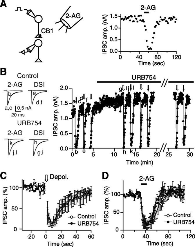
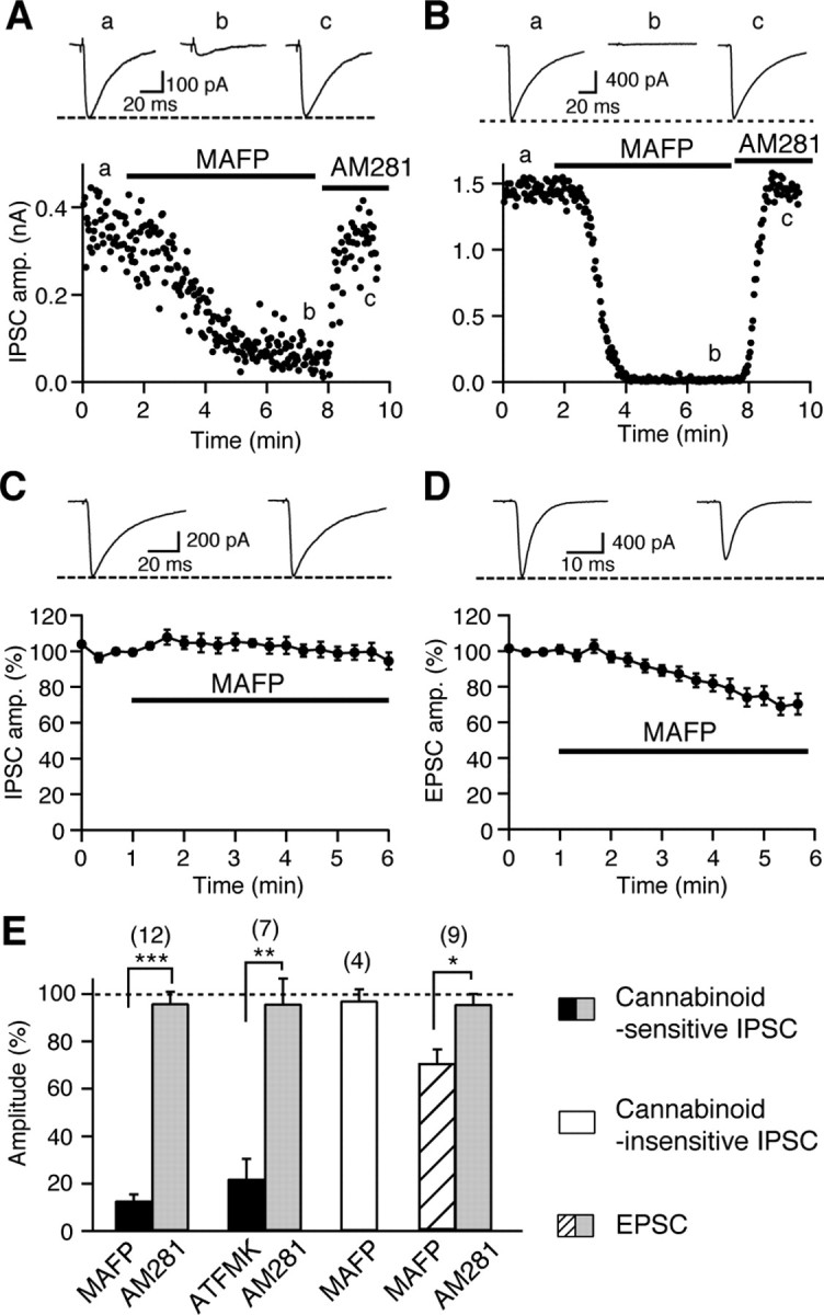
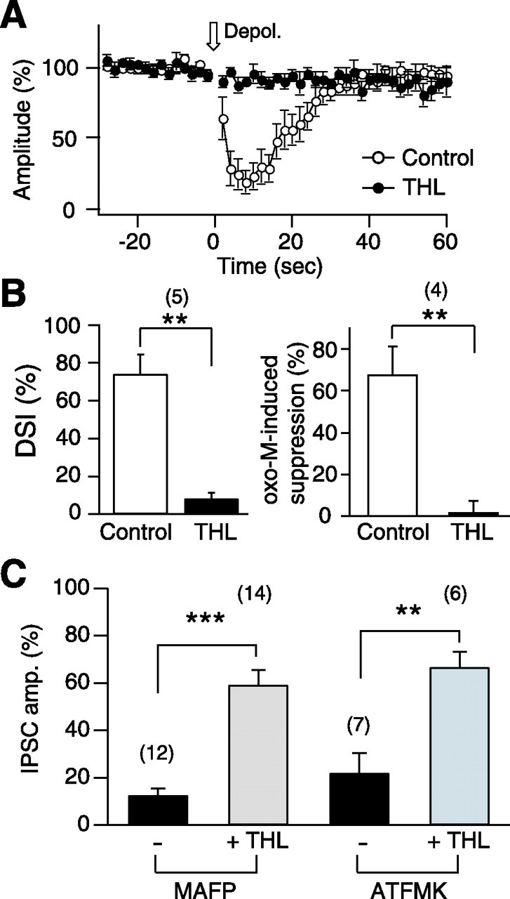
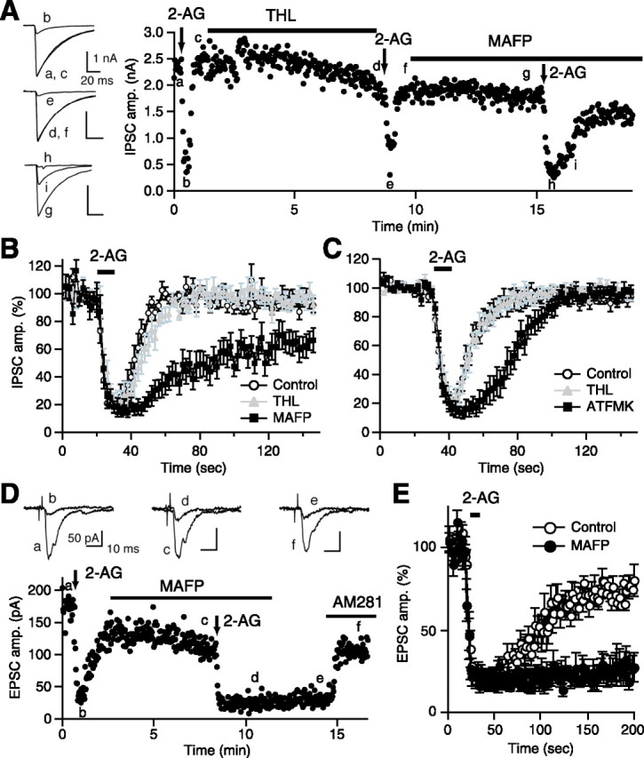
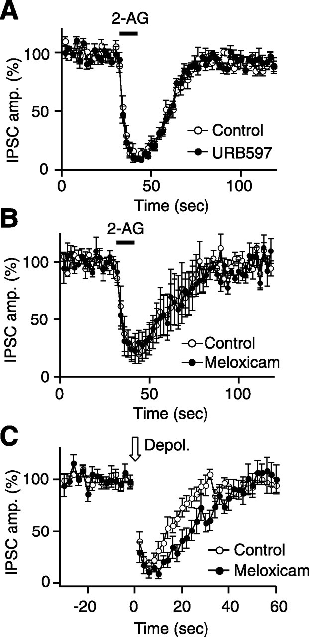
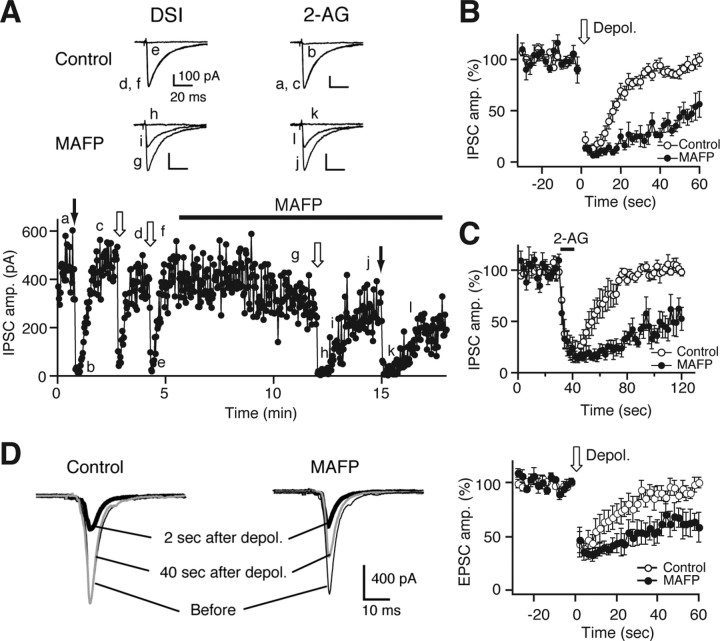
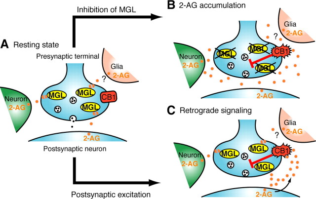
Endocannabinoids function as retrograde messengers and modulate synaptic transmission through presynaptic cannabinoid CB1 receptors. The magnitude and time course of endocannabinoid signaling are thought to depend on the balance between the production and degradation of endocannabinoids. The major endocannabinoid 2-arachidonoylglycerol (2-AG) is hydrolyzed by monoacylglycerol lipase (MGL), which is shown to be localized at axon terminals. In the present study, we investigated how MGL regulates endocannabinoid signaling and influences synaptic transmission in the hippocampus. We found that MGL inhibitors, methyl arachidonoyl fluorophosphonate and arachidonoyl trifluoromethylketone, caused a gradual suppression of cannabinoid-sensitive IPSCs in cultured hippocampal neurons. This suppression was reversed by blocking CB1 receptors and was attenuated by inhibiting 2-AG synthesis, indicating that MGL scavenges constitutively released 2-AG. We also found that the MGL inhibitors significantly prolonged the suppression of both IPSCs and EPSCs induced by exogenous 2-AG and depolarization-induced suppression of inhibition/excitation, a phenomenon known to be mediated by retrograde endocannabinoid signaling. In contrast, inhibitors of other endocannabinoid hydrolyzing enzymes, fatty acid amide hydrolase and cyclooxygenase-2, had no effect on the 2-AG-induced IPSC suppression. These results strongly suggest that presynaptic MGL not only hydrolyzes 2-AG released from activated postsynaptic neurons but also contributes to degradation of constitutively produced 2-AG and prevention of its accumulation around presynaptic terminals. Thus, the MGL activity determines basal endocannabinoid tone and terminates retrograde endocannabinoid signaling in the hippocampus.


Figures

Figure 1.
Figure 1.
URB754 has no effect on DSI and 2-AG-induced suppression of IPSCs. A, Schematic drawing showing local application of 2-AG to a pair of voltage-clamped neurons with cannabinoid-sensitive synapses (left). Right shows a representative experiment, in which locally applied 2-AG (0.1 μm for 10 s) reversibly decreased the amplitudes of cannabinoid-sensitive IPSCs. B, Examples of IPSC traces (left) and the time course of IPSC amplitude (right) from a representative experiment on the effects of URB754. DSI and 2-AG-induced suppression were induced repeatedly by postsynaptic depolarization (0 mV, 5 s; open arrows) and by local 2-AG application (0.1 μm, 10 s; filled arrows), respectively, before and during application of URB754 (5 μm). Sample IPSC traces (left) were acquired at the time points indicated in the graph (a–l, right). C, D, Averaged time courses of DSI (C) and 2-AG-induced suppression (D) obtained before and 5–10 min after the initiation of URB754 application (5 μm; n = 4).
Figure 2.
Figure 2.
MGL inhibitors specifically suppress cannabinoid-sensitive synaptic transmissions in a CB1-dependent manner. A, B, Two representative experiments showing the effects of bath-applied MAFP (0.1 μm) on cannabinoid-sensitive IPSCs. The IPSC traces acquired at the indicated time points (a–c) (top) and the IPSC amplitudes plotted as a function of time (bottom) are shown. The IPSC amplitude declined slowly (A) or rapidly (B) after MAFP application and recovered to the initial level after addition of the CB1 antagonist AM281 (0.3 μm). C, Effect of bath-applied MAFP (0.1 μm) on cannabinoid-insensitive IPSCs. Examples of IPSC traces before and 5 min after the initiation of MAFP application (top) and averaged data for the time course of IPSC amplitude (bottom; n = 4) are shown. D, Effects of bath-applied MAFP (0.1 μm) on EPSCs. Examples of EPSC traces before and 5 min after the initiation of MAFP application (top) and averaged data for the time course of EPSC amplitude (bottom; n = 9) are shown. E, From left to right, Averaged data showing percentage changes in the amplitudes of cannabinoid-sensitive IPSCs, cannabinoid-insensitive IPSCs, and EPSCs by application of indicated drugs. Numbers of tested cells are indicated in parentheses. *p < 0.05; **p < 0.01; ***p < 0.001 by paired t test.
Figure 3.
Figure 3.
The DAG lipase inhibitor THL eliminates endocannabinoid release and reduces IPSC suppression caused by MGL inhibitors. A, Blockade of DSI by THL. The graph shows averaged time course of DSI before and 5 min after bath application of 10 μm THL. B, Summary bar graphs showing the effects of THL on DSI (left) and endocannabinoid-mediated suppression of IPSCs by the muscarinic agonist oxo-M (3 μm). Oxo-M (3 μm) was bath applied for 1 min before (Control) and after (THL) treatment with THL (5 μm) for 5 min. C, Pretreatment with THL attenuates the suppression of IPSCs by MGL inhibitors. Effects of MAFP (0.1 μm) or ATFMK (10 μm) on cannabinoid-sensitive IPSCs were examined in the neuron pairs with (+THL) or without (−THL) pretreatment of THL (5 μm) for 5–7 min. Averaged data showing percentage changes in the amplitudes of cannabinoid-sensitive IPSCs 5 min after the initiation of MAFP or ATFMK application. Numbers of tested cells are indicated in parentheses. **p < 0.01; ***p < 0.001 by paired (A, B) or unpaired (C) t test.
Figure 4.
Figure 4.
MGL inhibitors prolong the 2-AG-induced suppression. A, A representative experiment showing effects of a sequential treatment with THL (5 μm) and MAFP (0.1 μm) on 2-AG-induced IPSC suppression. Downward arrows indicate the periods of 2-AG application. IPSC traces acquired at the indicated time points (a–i) are shown on the left. B, Average data for the time course of 2-AG-induced IPSC suppression before (Control) and after THL treatment and during additional treatment with MAFP (0.1 μm; n = 10). C, Average data for the time course of 2-AG-induced IPSC suppression before and after THL treatment and during additional treatment with ATFMK (10 μm; n = 6). D, E, An example (D) and the averaged data (E) showing effects of MAFP treatment on 2-AG-induced suppression of EPSCs. EPSC traces acquired at the indicated time points (a–f) are shown on the top. 2-AG (25 μm) was applied for 10 s (vertical arrows in D or a horizontal bar in E) before and after application of 0.1 μm MAFP. The 2-AG-induced persistent suppression of EPSC after MAFP application was reversed by addition of AM281 (0.3 μm). The averaged time courses of 2-AG-induced EPSC suppression (E) were obtained before and 5 min after the initiation of MAFP application (n = 5).
Figure 5.
Figure 5.
FAAH and COX-2 inhibitors have no effect on the time course of 2-AG-induced IPSC suppression. A, B, Average data for the time course of 2-AG-induced IPSC suppression obtained before and 5 min after the initiation of application of the FAAH inhibitor URB597 (1 μm) (A; n = 8) or the COX-2 inhibitor meloxicam (30 μm) (B; n = 4). C, Meloxicam slightly but significantly prolongs DSI. Average data for the time course of DSI before and after treatment with meloxicam for 5 min (30 μm; n = 8). The normalized IPSC amplitudes from 10 to 28 s after the depolarization were significantly smaller after the meloxicam treatment when compared with those before the treatment (p < 0.05 by paired t test).
Figure 6.
Figure 6.
The MGL inhibitor MAFP prolongs DSI, DSE, and 2-AG-induced IPSC suppression. A, Examples of IPSC traces (top) and the time course (bottom) of IPSC amplitude from a representative experiment. DSI and 2-AG-induced suppression were induced repeatedly by postsynaptic depolarization (0 mV, 5 s; open arrows) and by local 2-AG application (0.1 μm, 10 s; filled arrows), respectively, before and during application of MAFP (7 nm). IPSC traces were acquired at the indicated time points (a–l). B, C, Average data for the time course of DSI (B) and 2-AG-induced IPSC suppression (C) obtained before and 5–8 min after the initiation of 7 nm MAFP application (n = 6). D, Examples of EPSC traces (left) and the averaged time course of DSE (right; n = 4). Treatment with MAFP (7 nm) significantly prolonged DSE induced by 5 or 10 s postsynaptic depolarization of mouse hippocampal neurons. Thin, bold, and gray traces were acquired before, 2 s after, and 40 s after depolarization, respectively.
Figure 7.
Figure 7.
A model for the roles of presynaptic MGL in regulating basal endocannabinoid tone and terminating phasic endocannabinoid actions dependent on postsynaptic neuronal activity. A, At a resting state, a certain amount of 2-AG is constitutively produced and released from neurons or glial cells. The released 2-AG then diffuses into presynaptic terminal membranes and is rapidly inactivated by MGL so that CB1 receptors are not activated. B, When MGL is blocked at a resting state, 2-AG is accumulated around presynaptic terminals, and its local concentration is elevated high enough to tonically activate CB1 receptors. C, When a large amount of 2-AG is produced and released from activated postsynaptic neurons, the released 2-AG activates CB1 receptors, and the 2-AG signal is terminated by its degradation by presynaptic MGL.

Similar articles

Cited by

References

    1. Alger BE. Retrograde signaling in the regulation of synaptic transmission: focus on endocannabinoids. Prog Neurobiol. 2002;68:247–286. - PubMed
    1. Bisogno T, Howell F, Williams G, Minassi A, Cascio MG, Ligresti A, Matias I, Schiano-Moriello A, Paul P, Williams EJ, Gangadharan U, Hobbs C, Di Marzo V, Doherty P. Cloning of the first sn1-DAG lipases points to the spatial and temporal regulation of endocannabinoid signaling in the brain. J Cell Biol. 2003;163:463–468. - PMC - PubMed
    1. Chevaleyre V, Castillo PE. Heterosynaptic LTD of hippocampal GABAergic synapses: a novel role of endocannabinoids in regulating excitability. Neuron. 2003;38:461–472. - PubMed
    1. Chevaleyre V, Takahashi KA, Castillo PE. Endocannabinoid-mediated synaptic plasticity in the CNS. Annu Rev Neurosci. 2006;29:37–76. - PubMed
    1. Cravatt BF, Giang DK, Mayfield SP, Boger DL, Lerner RA, Gilula NB. Molecular characterization of an enzyme that degrades neuromodulatory fatty-acid amides. Nature. 1996;384:83–87. - PubMed

Publication types

Substances