. 2007 Feb 6;104(6):1835-40.
doi: 10.1073/pnas.0604893104. Epub 2007 Jan 31.

Lack of myostatin results in excessive muscle growth but impaired force generation

Affiliations

Lack of myostatin results in excessive muscle growth but impaired force generation

Helge Amthor et al. Proc Natl Acad Sci U S A. .

Erratum in

  • Proc Natl Acad Sci U S A. 2007 Mar 6;104(10):4240

Abstract

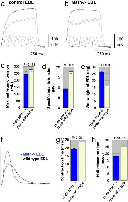
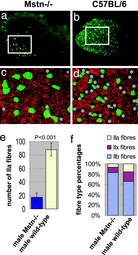
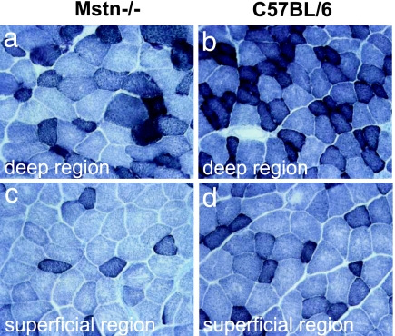
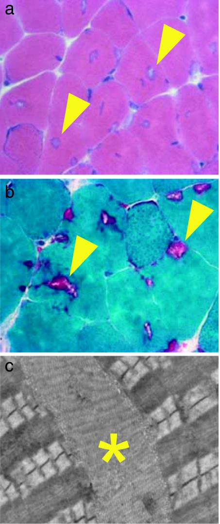
The lack of myostatin promotes growth of skeletal muscle, and blockade of its activity has been proposed as a treatment for various muscle-wasting disorders. Here, we have examined two independent mouse lines that harbor mutations in the myostatin gene, constitutive null (Mstn(-/-)) and compact (Berlin High Line, BEH(c/c)). We report that, despite a larger muscle mass relative to age-matched wild types, there was no increase in maximum tetanic force generation, but that when expressed as a function of muscle size (specific force), muscles of myostatin-deficient mice were weaker than wild-type muscles. In addition, Mstn(-/-) muscle contracted and relaxed faster during a single twitch and had a marked increase in the number of type IIb fibers relative to wild-type controls. This change was also accompanied by a significant increase in type IIB fibers containing tubular aggregates. Moreover, the ratio of mitochondrial DNA to nuclear DNA and mitochondria number were decreased in myostatin-deficient muscle, suggesting a mitochondrial depletion. Overall, our results suggest that lack of myostatin compromises force production in association with loss of oxidative characteristics of skeletal muscle.


Conflict of interest statement

The authors declare no conflict of interest.

Figures

Fig. 1.
Fig. 1.
Physiological properties and size of EDL muscles from male adult Mstn−/− mice compared with age-matched C57BL/6 wild types. (a and b) Records of superimposed isometric contraction in response to direct muscle stimulation at 1, 10, 20, 50, 100, and 200 Hz are shown for EDL muscles from wild-type (a) and Mstn−/− (b) mice. (c) Maximal Po of EDL muscles from Mstn−/− mice (blue column) compared with wild types (yellow column) (P = 0.189). (d) Maximal Po expressed as a function of muscle weight of EDL muscles from Mstn−/− mice (blue column) compared with wild types (yellow column) (P < 0.001). (e) Wet weight of EDL muscles that were measured after force testing from Mstn−/− mice (blue column) compared with wild types (yellow column) (P < 0.001). (f) Records of isometric twitch contraction of EDL muscles from a Mstn−/− mouse (blue trace) and a wild-type mouse (black trace). (g) Contraction times after a single twitch stimulation of EDL muscles from Mstn−/− mice (blue column) compared with wild types (yellow column) (P < 0.001). (h) Relaxation times after a single twitch stimulation of EDL muscles from Mstn−/− mice (blue column) compared with wild types (yellow column) (P < 0.001).
Fig. 2.
Fig. 2.
Muscle fiber-type profiles in EDL muscles from male Mstn−/−- and C57BL/6 wild-type mice. (a and b) Transverse sections of whole EDL (5× objective). The green fluorescence in a shows expression of MHC IIa in only a few fibers of Mstn−/− EDL compared with C57BL/6 wild-type EDL shown in b. IIa fibers can be seen to predominate in the deep muscle regions of the muscles, which are shown on the left side of the images. The white frames show areas of deep muscle region where the fiber type distribution was analyzed. The regions outlined in a and b are shown enlarged in c and d and show an overlay of staining for MHC IIa (green) and MHC IIb (red) by using ×20 objective. Asterisks depict non-IIa/non-IIb fibers. (e) Total number of IIa fibers in the whole EDL muscle sections from Mstn−/− mice (blue column) compared with wild types (yellow column) (P < 0.001). (f) Histogram showing the fiber-type distribution in the deep region of the EDL muscles. Fibers expressing MHC IIa and IIb and fibers negative for IIa/IIb were counted. Non-IIa/non-IIb fibers were considered as IIx fibers because there were hardly any fibers expressing slow MHC.
Fig. 3.
Fig. 3.
Oxidative properties of adult EDL muscles from 7-month-old male Mstn−/− mice relative to age-matched C57BL/6 wild types. SDH staining of Mstn−/− EDL (a and c) and of control EDL (b and d) in the deep (a and b, respectively) and superficial (c and d, respectively) regions. Darkly stained fibers contain high SDH activity, and pale-stained fibers contain low SDH activity.
Fig. 4.
Fig. 4.
Histological analysis of tubular aggregates from adult EDL muscles of Mstn−/− mice. (a) Transverse sections of the EDL muscles from a 7-month-old male Mstn−/− mouse after staining with H&E showing the presence of numerous cytoplasmic inclusions (arrowheads) in many muscle fibers. (b) Transverse sections of the EDL from a 7-month-old male Mstn−/− mouse after Gomori's trichrome staining showing cytoplasmic inclusions in purple color (arrowheads). (c) Electron micrograph taken from an EDL of a 7-month-old male Mstn−/− mouse showing stacks of tubular and saccular formations (∗), which define tubular aggregates, between the contractile apparatus.

References

    1. McPherron AC, Lawler AM, Lee SJ. Nature. 1997;387:83–90. - PubMed
    1. Lee SJ. Annu Rev Cell Dev Biol. 2004;20:61–86. - PubMed
    1. Schuelke M, Wagner KR, Stolz LE, Hubner C, Riebel T, Komen W, Braun T, Tobin JF, Lee SJ. N Engl J Med. 2004;350:2682–2688. - PubMed
    1. Ferrell RE, Conte V, Lawrence EC, Roth SM, Hagberg JM, Hurley BF. Genomics. 1999;62:203–207. - PubMed
    1. Wagner KR, McPherron AC, Winik N, Lee SJ. Ann Neurol. 2002;52:832–836. - PubMed

Publication types