. 2007 Apr;45(4):1225-33.
doi: 10.1128/JCM.02199-06. Epub 2007 Jan 31.

Discovery of a new capsular serotype (6C) within serogroup 6 of Streptococcus pneumoniae

Affiliations

Discovery of a new capsular serotype (6C) within serogroup 6 of Streptococcus pneumoniae

In Ho Park et al. J Clin Microbiol. 2007 Apr.

Abstract

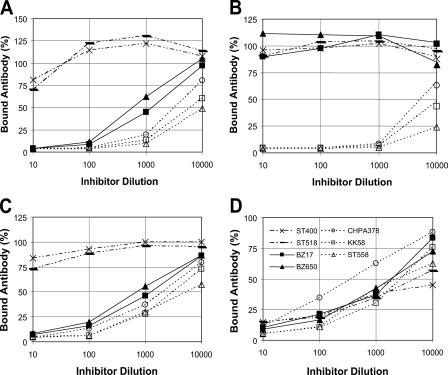
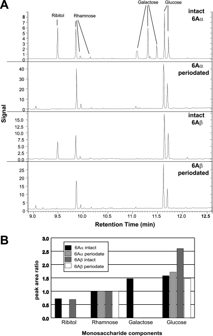
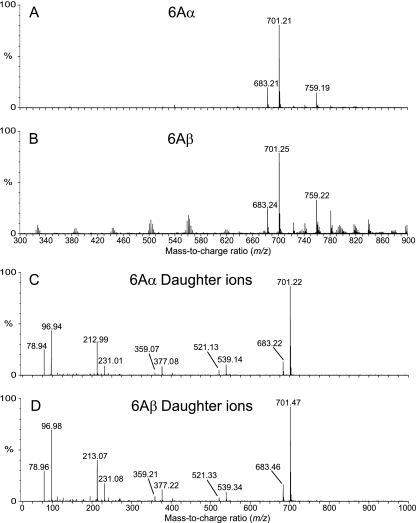
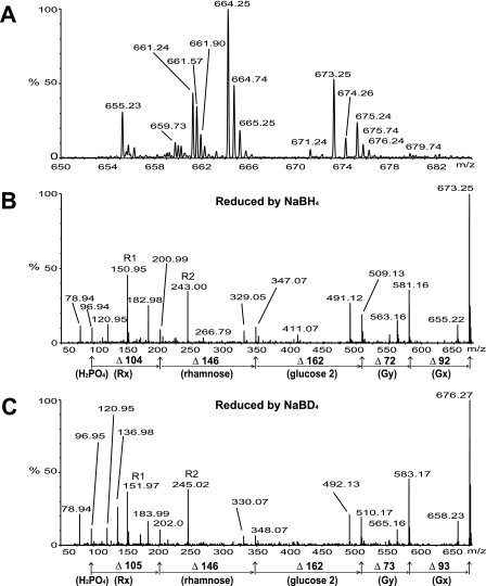
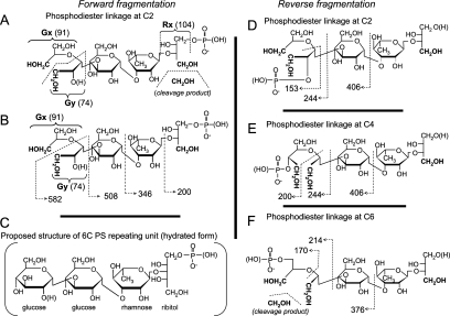
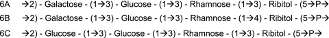
Using two monoclonal antibodies, we found subtypes among pneumococcal isolates that are typed as serotype 6A by the quellung reaction. The prevalent subtype bound to both monoclonal antibodies and was labeled here 6Aalpha, whereas the minor subtype bound to only one monoclonal antibody and was labeled 6Abeta. To determine the biochemical nature of the two serologically defined subtypes, we purified capsular polysaccharides (PSs) from the two subtypes and examined their chemical structures with gas-liquid chromatography and mass spectrometry. The study results for 6Aalpha PS are consistent with the previously published structure of 6A PS, which is -->2) galactose (1-->3) glucose (1-->3) rhamnose (1-->3) ribitol (5-->phosphate. In contrast, the 6Abeta PS study results show that its repeating unit is -->2) glucose 1 (1-->3) glucose 2 (1-->3) rhamnose (1-->3) ribitol (5-->phosphate. We propose to continue referring to 6Aalpha as serotype 6A but to refer to 6Abeta as serotype 6C. Serotype 6C would thus represent the 91st pneumococcal serotype, with 90 pneumococcal serotypes having previously been recognized. This study also demonstrates that a new serotype may exist within an established and well-characterized serogroup or serotype.


Figures

FIG. 1.
FIG. 1.
Antibody bound to ELISA plates (y axis) against the dilution of pneumococcal lysates (x axis). Lysates include two 6Aβ isolates (solid symbols with continuous lines), three 6Aα isolates (open symbols with dotted lines), and two 6B isolates (dashed connecting lines). Antibodies used for the assay were Hyp6AG1 (A), Hyp6AM3 (B), rabbit serum pool Q (C), and rabbit “factor 6b” serum (D).
FIG. 2.
FIG. 2.
Carbohydrate composition (A) of capsular PS from 6Aα (top two graphs) and 6Aβ (bottom two graphs) before and after periodate treatment. The monosaccharides are identified in the top chromatogram. B shows normalized peak areas of each monosaccharide for 6Aα and 6Aβ. The peak areas of all monosaccharides from each PS are normalized to the peak area of the associated rhamnose. 6Aβ shows no galactose peaks but has about twice as much glucose as 6Aα does.
FIG. 3.
FIG. 3.
Mass spectra of the repeating units of 6Aα (A) and 6Aβ (B) and their daughter ions (C and D, respectively). The mass-to-charge ratio (m/z) was rounded off to two decimal points.
FIG. 4.
FIG. 4.
Mass spectrum of the repeating unit of 6Aβ PS after oxidation and reduction (A) and daughter ions (B and C). The sample used for B was reduced with NaBH4, and that used for C was reduced with NaBD4. The mass-to-charge ratio (m/z) was rounded off to two decimal points. R1 and R2 (in B and C) indicate that the peaks corresponding to ions derived by reverse fragmentations. Numbers following the delta symbol indicate the m/z unit differences between the peaks and associated with the names of the fragments. All the peaks in C correspond to the peaks in B except for a peak at 136.98, which may be a contaminant.
FIG. 5.
FIG. 5.
Proposed chemical structures of 6C capsular polysaccharides and the structure of its cleavage products. The proposed structure of the 6C repeating unit is shown in C. A and B show possible molecular ions if the phosphate group is attached to ribitol and if the phosphodiester is linked to the second carbon of glucose 1. D, E, and F indicate potential cleavage patterns of the repeating unit if the phosphodiester is linked to the second (D), the fourth (E), or the sixth (F) carbon of glucose 1. Hydrated forms are shown, and the residues involved in hydration are shown in parentheses. Periodate-sensitive sites are shown in boldface type, and cleavage products are shown in A and F. Potential molecular ions are shown with dotted lines with arrows along with their AMUs. Gx and Gy are potential glucose 1 fragments, and Rx is the remaining ribitol fragment after oxidation and reduction reactions. Their AMUs are shown in parenthesis.
FIG. 6.
FIG. 6.
Comparison of 6A, 6B, and 6C PS structures. 6B PS differs from 6A PS in its rhamnose-ribitol linkage. 6C PS differs from 6A PS by having a glucose residue in place of a galactose residue.

Similar articles

Cited by

References

    1. Abeygunawardana, C., T. C. Williams, J. S. Sumner, and J. P. Hennessey, Jr. 2000. Development and validation of an NMR-based identity assay for bacterial polysaccharides. Anal. Biochem. 279:226-240. - PubMed
    1. Avery, O. T., and R. Dubos. 1931. The protective action of a specific enzyme against type III pneumococcus infection in mice. J. Exp. Med. 54:73-89. - PMC - PubMed
    1. Butler, J. C., R. F. Breiman, J. F. Campbell, H. B. Lipman, C. V. Broome, and R. R. Facklam. 1993. Pneumococcal polysaccharide vaccine efficacy. An evaluation of current recommendations. JAMA 270:1826-1831. - PubMed
    1. Cole, R. 1913. Treatment of pneumonia by means of specific serums. JAMA 61:663-666.
    1. Cole, R. B. 1997. Electrospray ionization mass spectrometry: fundamentals, instrumentation, and applications. Wiley, New York, NY.

Publication types

MeSH terms

Substances

LinkOut - more resources