. 2007 Apr;18(4):1282-92.
doi: 10.1091/mbc.e06-09-0846. Epub 2007 Jan 31.

Subnuclear localization and dynamics of the Pre-mRNA 3' end processing factor mammalian cleavage factor I 68-kDa subunit

Affiliations

Subnuclear localization and dynamics of the Pre-mRNA 3' end processing factor mammalian cleavage factor I 68-kDa subunit

Stefano Cardinale et al. Mol Biol Cell. 2007 Apr.

Abstract

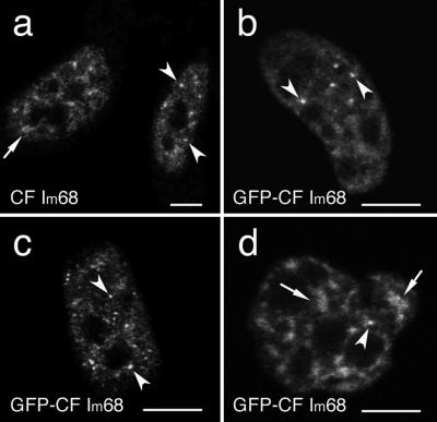
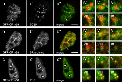
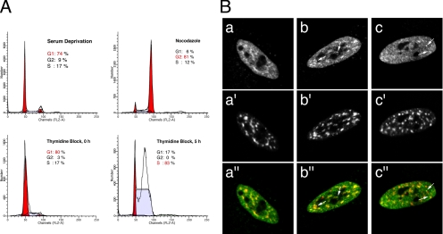
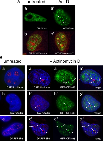
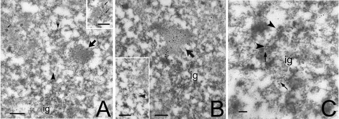
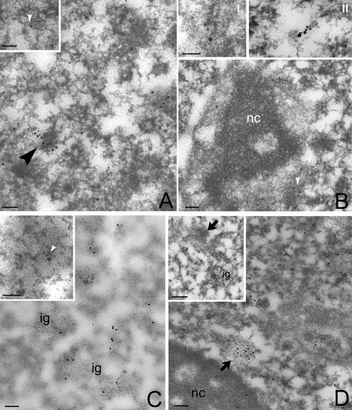
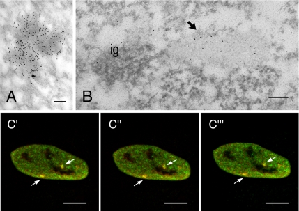
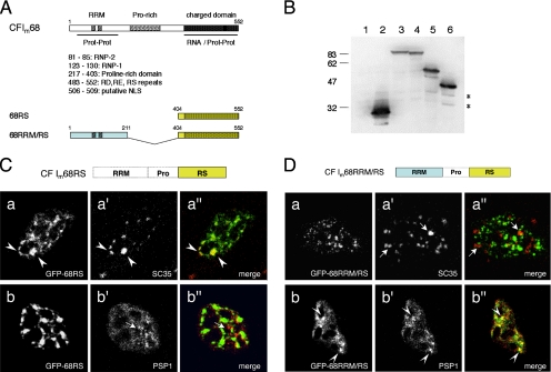
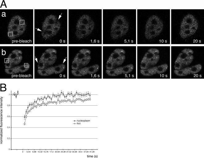
Mammalian cleavage factor I (CF Im) is an essential factor that is required for the first step in pre-mRNA 3' end processing. Here, we characterize CF Im68 subnuclear distribution and mobility. Fluorescence microscopy reveals that in addition to paraspeckles CF Im68 accumulates in structures that partially overlap with nuclear speckles. Analysis of synchronized cells shows that CF Im68 distribution in speckles and paraspeckles varies during the cell cycle. At an ultrastructural level, CF Im68 is associated with perichromatin fibrils, the sites of active transcription, and concentrates in interchromatin granules-associated zones. We show that CFIm68 colocalizes with bromouridine, RNA polymerase II, and the splicing factor SC35. On inhibition of transcription, endogenous CF Im68 no longer associates with perichromatin fibrils, but it can still be detected in interchromatin granules-associated zones. These observations support the idea that not only splicing but also 3' end processing occurs cotranscriptionally. Finally, fluorescence recovery after photobleaching analysis reveals that the CF Im68 fraction associated with paraspeckles moves at a rate similar to the more dispersed molecules in the nucleoplasm, demonstrating the dynamic nature of this compartment. These findings suggest that paraspeckles are a functional compartment involved in RNA metabolism in the cell nucleus.


Figures

Figure 1.
Figure 1.
CF Im 68-kDa subunit is concentrated in speckle-like structures as well as in discrete foci. HeLa cells were either immunolabeled with a polyclonal antibody directed against CF Im68 (a) or transiently transfected with GFP-CF Im68 (b–d). Confocal microscope sections show that CF Im68 is detected in the nucleus, excluding the nucleoli; the protein is diffused in the nucleoplasm with additional concentration in speckled enlargements (broken arrows) and in bright foci (arrowheads). Bar, 10 μm.
Figure 2.
Figure 2.
Interconnection of CF Im68 speckles and nuclear speckles. Confocal microscope sections of HeLa cells that were transiently transfected with pEGFP-CF Im68, and subsequently immunostained with antibodies against SC35 (a′), SR proteins (b′), or PSP1 (c′). The images of the green fluorescence of pEGFP-CF Im68 (a–c) and the red fluorescence of the antibodies are merged in a″, b″, and c″. Three sequential planes of two different high magnification images (IV–VI, and VII–IX) that were taken from different cells demonstrate the degree of overlap between different compartments. Broken arrows indicate CF Im68 foci. Arrowheads indicate colocalization in paraspeckles. Bar, 10 μm.
Figure 3.
Figure 3.
CF Im68 localization is regulated during the cell cycle. HeLa cells were arrested in G0 by serum deprivation, at the G2/M phase using nocodazole, or at the G1/S transition using the double thymidine block procedure (as indicated in A). Cells were then fixed and used for fluorescence-activated cell sorting analysis or double-stained with anti-CF Im68 antiserum as described in Materials and Methods (B, a–c) and anti-SC35 antibody (B, a′–c′). After serum deprivation, CF Im68 concentrated in speckles in 64% of the cells (B, a–a″). In 55% of the cells arrested at G2/M, CF Im68 localized in speckles as well as in one to three foci (B, b–b″). At the G1/S transition and in S-phase CF Im68 was concentrated in foci in ∼60% of the cells (B, c–c″). Quantification of the cellular localization pattern was performed as described in Materials and Methods. The images of the green fluorescence of CF Im68 and the red fluorescence of SC35 are merged in panels a″, b″, and c″. Broken arrows indicate foci.
Figure 4.
Figure 4.
Inhibition of transcription results in the relocalization of CF Im68 to perinucleolar caps. (A) CF Im68 relocalizes in cap structures upon inhibition of transcription. Confocal microscope sections through HeLa cells transiently transfected with GFP-CF Im68 (green) either untreated (a–b) or incubated with 5 μg/ml actinomycin D for 2 h (a′–b′). Cells were stained with pironin Y to show cellular RNA, including nucleoli (red, b and b′). Broken arrows indicate the perinucleolar cap-like structures. (B) CF Im68 colocalizes with PSP1 also after transcription inhibition. Fluorescence micrographs of sections through HeLa cells transiently transfected with GFP-CFIm68 (green) either untreated (a–c) or incubated with 5 μg/ml actinomycin D for 2 h (a′–c‴). Cells were then stained with DAPI to show DNA (blue). Cells were double labeled with antibodies to fibrillarin (red, a and a″), to p80 coilin (red, b and b″), or to PSP1 (red, c and c″). Broken arrows indicate relocalized GFP-CF Im68 at the nucleolar periphery after treatment with actinomycin D; arrowheads indicate relocalized fibrillarin (a and a‴), coilin (b and b‴), and PSP1 (c and c‴), respectively. The sections were obtained using the DeltaVision system, and the images were deconvolved using softWoRx (Applied Precision).
Figure 5.
Figure 5.
Ultrastructural localization of CF Im68. HeLa cells were embedded in LR White resin, sectioned, and immunogold labeled with a polyclonal antiserum that recognizes CF Im large subunits (Rüegsegger et al., 1998). Bar, 0.2 μm. (A) At low magnification, a large zone labeled for CF Im (large arrow) is present nearby a cluster of interchromatin granules (ig). The labeling is visible also on perichromatin granules (small arrows) and on PFs (arrowheads). Inset, group of labeled PGs. (B) At higher magnification, the labeled area (large arrow) is recognizable as an IGAZ and is associated with ig. Inset, a single PF is labeled by CFIm. (arrowhead). (C) Double immunolabeling for CFIm (large grains, arrowheads) and SC35 (small grains, broken arrows). Labeling for SC35 is present on the IG cluster, whereas CFIm mostly localizes at the periphery of the IGCs.
Figure 6.
Figure 6.
CF Im colocalizes with nascent transcripts. (A) RNA polymerase II (small grains) colocalizes with CFIm (large grains) on some PFs (arrowhead). Inset I, at higher magnification, the double labeling is shown on a single, nascent PF. Bar, 0.1 μm. (B) Newly incorporated BrU (small grains) colocalizes with CFIm (large grains) on PFs (arrowhead). Inset, a single, double-labeled PF. Nc, nucleolus. Inset II, a single PF, labeled with anti-BrU antibody (6-nm gold), anti-CF Im68 antiserum (12-nm gold), and anti-CstF antibody (18-nm gold). Bar, 0.1 μm. (C) Newly incorporated BrU (small grains) colocalizes with CFIm (large grains) on PFs (arrowhead). Inset, a single, double-labeled PF. Nc, nucleolus. Bar, 0.1 μm. (D) Immunolabeling for CFIm after selective staining of RNA with terbium. Inset, double immunolabeling for CFIm (large grains) and SC35 (small grains) of terbium-stained RNA. The arrowhead indicates labeling on PFs, whereas interchromatin granules (ig) are unlabeled. Bar, 0.1 μm. (E) HeLa cells treated with DRB. The labeling is decreased on PFs, but still intense on IGAZs (large arrow). Inset, another IGAZ labeled for CFIm after DRB treatment. Ig, interchromatin granules; nc, nucleolus. Bar, 0.1 μm.
Figure 7.
Figure 7.
CF Im68 and PSF colocalize in IGAZs and paraspeckles. (A) Immunolabeling for PSF concentrates on dense fibrillar areas. Bar, 0.1 μm. (B) HeLa cells were double labeled with a rabbit antiserum that recognizes CF Im68 and with mouse mAb that recognizes PSF. PSF (small grains) colocalizes with CFIm (large grains) on IGAZ (arrow) nearby a cluster of interchromatin granules (ig). Bar, 0.1 μm. (C) Fluorescence micrographs of three sequential planes sections through HeLa cells that were double labeled for CF Im68 and for PSF. The images of the green fluorescence of CF Im68 and the red fluorescence of PSF are merged in c′–c‴. Broken arrows indicate colocalization in paraspeckles. Bar, 10 μm.
Figure 8.
Figure 8.
CF Im68 deletions mutants identify regions important for the localization in speckles and in paraspeckles. (A) Domain structure of CF Im 68-kDa subunit. CF Im68 contains an N-terminal RNA recognition motif of the RNP type (RRM, amino acids [aa] 81-130), a central proline-rich region (Prorich, aa 217-403), and a C-terminal charged domain similar to the RS domain of SR proteins (aa 404-552) with a putative NLS (black box, aa 506-509). (B) Western blot analysis verifies the expression of GFP-fusion constructs in HeLa cells. Lane 1 is the nontransfected control lysate, lane 2 is GFP (expected molecular weight [MW] 27), lane 3 is GFP-CF Im68 (expected MW 86.1), lane 4 is GFP-68RRM/RS (expected MW 67), lane 5 is GFP-68RRM (expected MW 50.4), and lane 6 is GFP-68RS (expected MW 44.4). (C) The RS-like domain is sufficient for localization in speckles. In the left column are GFP images of HeLa cells transiently transfected with the RS fusion construct. The middle column shows fluorescence images of cells stained with antibodies that recognize either SC35 (a′) or PSP1 (b′). The right column shows the overlays of the GFP images (green) and the antibodies (red). Arrowheads indicate areas of colocalization (yellow). Broken arrows indicate paraspeckles. (D) The RRM is necessary for paraspeckles localization. In the left column are GFP images of HeLa cells after transient transfection of the RRM/RS fusion construct. The middle column shows fluorescence images of cells stained with antibodies that recognize either SC 35 (a′) or PSP1 (b′). The right column shows the overlays of the GFP images (green) and the antibodies (red). Arrowheads indicate areas of colocalization (yellow). Broken arrows indicate SC35 speckles.
Figure 9.
Figure 9.
Recovery of GFP-CF Im68 fluorescence after photobleaching in living cells. (A) Cells expressing GFP-CF Im68 were imaged before or after bleaching of a nucleoplasmic area (top row) or of paraspeckles (bottom row). Images were taken at the indicated times after the bleach pulse. The area of the bleach spot is indicated with a square. a, FRAP of nucleoplasmic regions. b, FRAP of paraspeckles. Bar, 5 μm. (B) Kinetics of recovery after spot bleaching of a nucleoplasmic region (closed circles) and of a paraspeckle (open diamonds). Recovery curves are plotted as normalized fluorescence intensity versus time. Each point is the mean value from eight cells. The error bars indicate the SD.

References

    1. Almeida F., Saffrich R., Ansorge W., Carmo-Fonseca M. Microinjection of anti-coilin antibodies affects the structure of coiled bodies. J. Cell Biol. 1998;142:899–912. - PMC - PubMed
    1. Andersen J. S., Lyon C. E., Fox A. H., Leung A. K., Lam Y. W., Steen H., Mann M., Lamond A. I. Directed proteomic analysis of the human nucleolus. Curr. Biol. 2002;12:1–11. - PubMed
    1. Bentley D. The mRNA assembly line: transcription and processing machines in the same factory. Curr. Opin. Cell Biol. 2002;14:336–342. - PubMed
    1. Bernhard W. A new staining procedure for electron microscopical cytology. J. Ultrastruct. Res. 1969;27:250–265. - PubMed
    1. Biggiogera M., Fakan S. Fine structural specific visualization of RNA on ultrathin sections. J. Histochem. Cytochem. 1998;46:389–395. - PubMed

Publication types

MeSH terms