Comparative Study
. 2007 Feb 21;26(4):1024-34.
doi: 10.1038/sj.emboj.7601562. Epub 2007 Feb 1.

Reconstitution and subunit geometry of human condensin complexes

Affiliations
Comparative Study

Reconstitution and subunit geometry of human condensin complexes

Itay Onn et al. EMBO J. .

Abstract

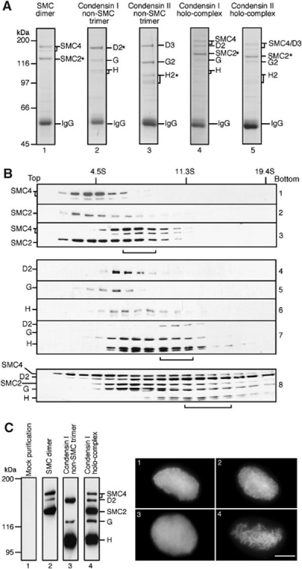
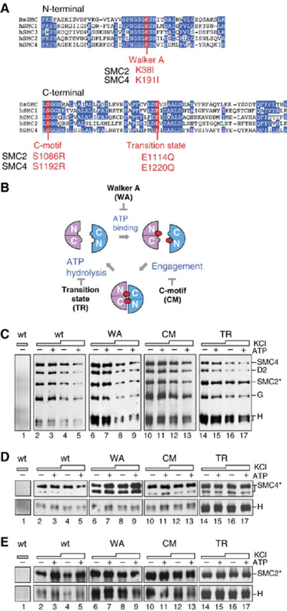
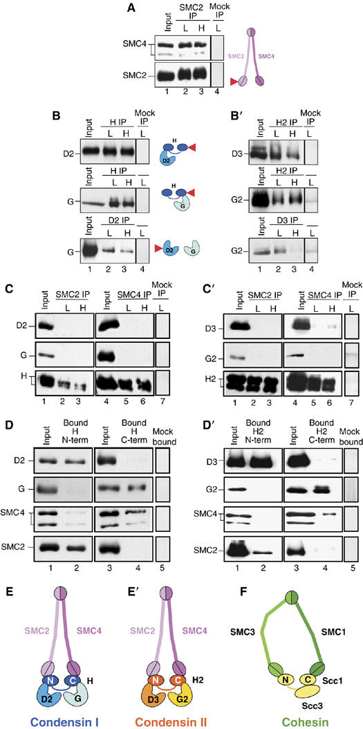
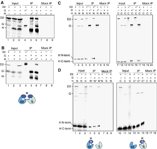
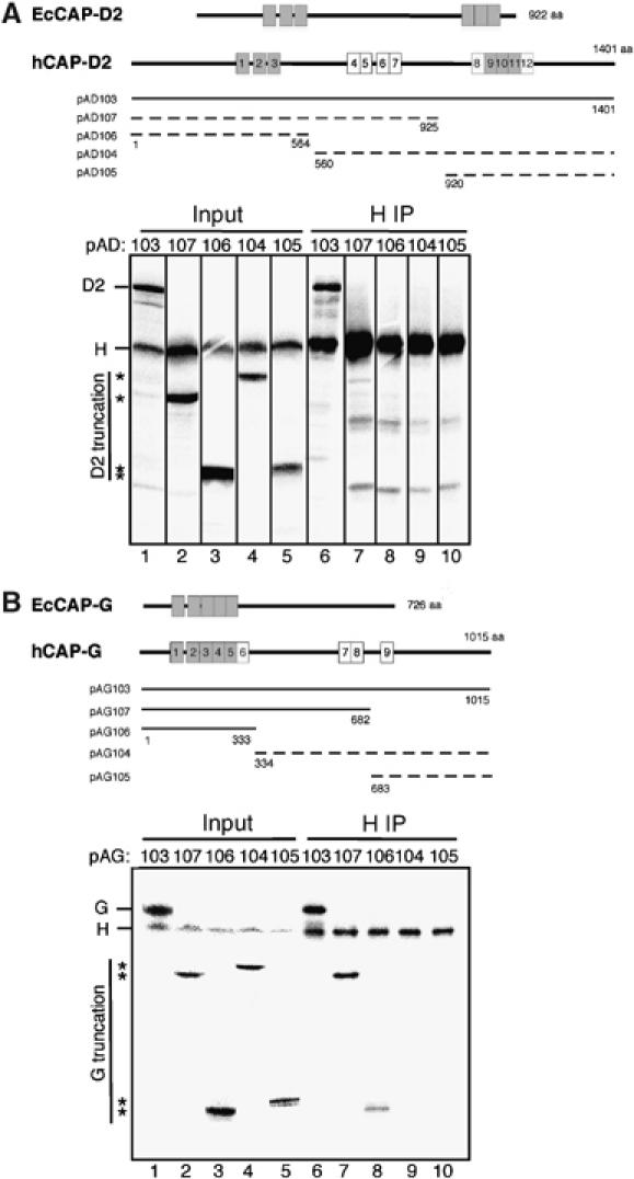
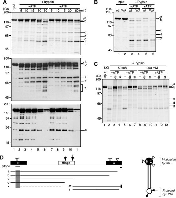
Vertebrate cells possess two different condensin complexes, known as condensin I and condensin II, that play a fundamental role in chromosome assembly and segregation during mitosis. Each complex contains a pair of structural maintenance of chromosomes (SMC) ATPases, a kleisin subunit and two HEAT-repeat subunits. Here we use recombinant human condensin subunits to determine their geometry within each complex. We show that both condensin I and condensin II have a pseudo-symmetrical structure, in which the N-terminal half of kleisin links the first HEAT subunit to SMC2, whereas its C-terminal half links the second HEAT subunit to SMC4. No direct interactions are detectable between the SMC dimer and the HEAT subunits, indicating that the kleisin subunit acts as the linchpin in holocomplex assembly. ATP has little, if any, effects on the assembly and integrity of condensin. Cleavage pattern of SMC2 by limited proteolysis is changed upon its binding to ATP or DNA. Our results shed new light on the architecture and dynamics of this highly elaborate machinery designed for chromosome assembly.


Figures

Figure 1
Figure 1
Characterization of recombinant condensin subunits and complexes. (A) Subunits of the condensin complexes were coexpressed in different combinations in Sf9 cells and the cell lysate was subjected to immunoprecipitation using an antibody specific to one of the coexpressed subunits (indicated by asterisks). The sub- and holocomplexes reconstituted were the SMC dimer (hSMC2 and hSMC4; lane 1), the non-SMC trimer of condensin I (hCAP-D2, hCAP-G and hCAP-H; lane 2), the non-SMC trimer of condensin II (hCAP-D3, hCAP-G2 and hCAP-H2; lane 3), the holocomplex of condensin I (lane 4) and the holocomplex of condensin II (lane 5). Proteins were resolved by 7.5% SDS–PAGE and stained with Coomassie brilliant blue (CBB). A very low background was observed as judged by CBB staining or immunoblotting when an untrasnfected Sf9 extract was subjected to immunoprecipitation using these antibodies (data not shown). (B) Lysates of Sf9 cells expressing individual condensin subunits or coexpressing subunits in different combinations were loaded onto 5–20% sucrose gradients and centrifuged at 36 000 r.p.m. for 15 h in an SW50.1 rotor (Beckman). Fractions were precipitated with trichloroacetic acid, resolved by 7.5% SDS–PAGE and analyzed by immunoblotting with the indicated antibodies. The positions of three protein standards (BSA (4.5S), catalase (11.3S) and thyroglobulin (19.4S)) are indicated. The peak fractions of reconstituted complexes are indicated by brackets: the 8S SMC dimer (panel 3); the 11S non-SMC trimer (panel 7); the 13S holocomplex (panel 8). (C) Fractions enriched with the hSMC2–hSMC4 dimer (lane 2), the non-SMC trimer of condensin I (lane 3) and the holocomplex of condensin I (lane 4) were prepared by single-step affinity chromatography and analyzed by immunoblotting. Mock purification was performed using an untransfected cell lysate (lane 1). A Xenopus egg HSS depleted of endogeneous condensin I was supplemented with the mock-purified fraction (panel 1), the SMC dimer fraction (panel 2), the non-SMC trimer fraction (panel 3) or the holocomplex fraction (panel 4), and the ability of each mixture to convert sperm chromatin into mitotic chromosome-like structures was assayed microscopically. Bar, 10 μm.
Figure 2
Figure 2
ATP is not required for holocomplex assembly or SMC–kleisin interactions. (A) Sequence alignment of the N- and C-terminal conserved domains among the human SMC proteins (hSMC1–4) and the Bacillus subtilis SMC protein (BsSMC). Also indicated are the mutation sites introduced into hSMC2 and hSMC4 in the current study. (B) The proposed SMC ATPase cycle. Each head domain is composed of the N- and C-terminal domains of an SMC subunit. Binding of ATP (closed circles) to the head domains induces their engagement, and hydrolysis of ATP triggers their disengagement. The Walker A mutation (WA) prevents ATP binding, whereas the C-motif mutation (CM) blocks engagement. The transition-state mutation (TR) stabilizes engagement by slowing down ATP hydrolysis. (C) A pair of wild-type or mutant SMC subunits was coexpressed with the three non-SMC subunits of condensin I in Sf9 cells. The lysates were subjected to immunoprecipitation using a non-immune IgG (lane 1) or anti-hSMC2 (lanes 2–17), and washed with a buffer containing 0.1 M KCl (lanes 1, 2, 3, 6, 7, 10, 11, 14 and 15) or 0.5 M KCl (lanes 4, 5, 8, 9, 12, 13, 16 and 17). No ATP (−) or ATP (+) was added in the lysate and the washing buffers throughout the procedures. The precipitates were analyzed by immunoblotting using the indicated antibodies. When mutant complexes were assayed, both hSMC2 and hSMC4 subunits contained the corresponding mutations. (D) hCAP-H was coexpressed with the wild-type or mutant forms of hSMC4, immunoprecipitated with a non-immune IgG (lane 1) or anti-hSMC4 (lanes 2–17) and analyzed as above. (E) Similarly, hCAP-H was coexpressed with the wild-type or mutant forms of hSMC2 and immunoprecipitated with a non-immune IgG (lane 1) or anti-hSMC2 (lanes 2–17).
Figure 3
Figure 3
Pseudo-symmetrical structure of condensin I and condensin II as revealed by subunit-subunit interaction assays. (A) A lysate was prepared from Sf9 cells coexpressing hSMC2 and hSMC4 (lane 1) and was subjected to immunoprecipitation with anti-hSMC2 (arrow). The precipitates were recovered on protein A beads, washed with a buffer containing 0.1 M (L, lane 2) or 0.5 M KCl (H, lane 3) and analyzed by immunoblotting with anti-hSMC4 and anti-hSMC2. (B) Two of the three non-SMC subunits of condensin I were coexpressed in different combinations, immunoprecipitated with the indicated antibodies (arrows) and analyzed as above. In (A) and (B), subunit–subunit interactions detected are summarized in the adjacent cartoons. Target subunits used for immunoprecipitations are indicated by the arrows. Mock immunoprecipitation using non-immune rabbit IgG is shown in lane 4 of each panel. (C) hSMC2 was coexpressed with one of the three non-SMC subunits of condensin I, immunoprecipitated with anti-hSMC2 and analyzed as above (left panel, lanes 1–3). The same set of experiments was performed for hSMC4 (right panel, lanes 4–6). A mock immunoprecipitation is shown in lane 7. (D) A hexahistidine-tagged, N-terminal fragment of hCAP-H was coexpressed with either hCAP-D2, hCAP-G, hSMC4 or hSMC2 in Sf9 cells. Lysates were prepared and mixed with Ni-NTA beads. After washing the beads with a buffer containing 0.1 M KCl, bound proteins were analyzed by immunoblotting with the indicated antibodies (left). The same set of experiments was performed for a hexahistidine-tagged, C-terminal fragment of hCAP-H (right). A mock pull-down is shown in lane 5. (E) Deduced subunit geometry in the condensin I complex. (B′–D′) An identical set of experiments was performed as in (B–D) by using condensin II subunits. (E′) Deduced subunit geometry in the condensin II complex. (F) Subunit geometry in the cohesin complex proposed by Haering et al (2002).
Figure 4
Figure 4
hCAP-D2 and hCAP-G bind independently to separate domains of hCAP-H. (A) Full-length hCAP-D2, hCAP-G and hCAP-H were translated in vitro in the presence of [35S]methionine. The reactions were mixed in different combinations (input; lanes 1–3) and subjected to immunoprecipitation with anti-hCAP-G (lane 4), anti-hCAP-H (lanes 5 and 6) or non-immune rabbit IgG (lanes 7–9). The input and bound fractions were fractionated by SDS–PAGE and analyzed by autoradiography. (B) Full-length hCAP-D2, hCAP-G and hCAP-H were translated in vitro in the presence of [35S]methionine. The reactions were mixed in different combinations (input; lanes 1–3) and subjected to immunoprecipitation with anti-hCAP-D2 (lane 4), anti-hCAP-H (lanes 5 and 6) or non-immune IgG (lanes 7–9). (C) An N-terminal fragment of hCAP-H (1-393), full-length hCAP-D2 and hCAP-G were translated in vitro in the presence of [35S]methionine. The reactions were mixed in different combinations as indicated (input; lanes 1–3), and subjected to immunoprecipitation with anti-hCAP-D2 (lanes 4 and 6), anti-hCAP-G (lanes 5 and 7) or non-immune IgG (lanes 8–10). Alternatively, the N-terminal fragment was replaced with a C-terminal fragment of hCAP-H (395–730), and a similar set of experiments was performed (lanes 11–20). (D) An N-terminal fragment of hCAP-H (1-393), full-length hCAP-D2 and hCAP-G were translated in vitro in the presence of [35S]methionine. The reactions were mixed in different combinations as indicated (input; lanes 1–3) and subjected to immunoprecipitation with the Penta-His antibody (Qiagen) that recognizes the N-terminal fragment of hCAP-H (lanes 4–6) or non-immune rabbit IgG (lanes 7–9). Alternatively, the N-terminal fragment was replaced with a C-terminal fragment of hCAP-H (395–730), and anti-hCAP-H that recognizes its C-terminal fragment or non-immune IgG was used for immunoprecipitation (lanes 10–18). In (A–D), target subunits used for immunoprecipitations are indicated by the asterisks.
Figure 5
Figure 5
Domain dissection of the HEAT subunits. (A) (upper) Overall structures of hCAP-D2 and its ortholog in Encephalitozoon cuniculi (EcCAP-D2) are shown (upper). Among the 12 HEAT repeats found in hCAP-D2 (shown by boxes), only six are detectable in EcCAP-D2 (gray boxes); (lower) a deletion series of hCAP-D2 (pAD103–107) was translated in vitro and each reaction was mixed with a reaction containing full-length hCAP-H (input; lanes 1–5). The mixtures were subjected to immunoprecipitation with anti-hCAP-H (IP; lanes 6–10). The input and precipitated fractions were fractionated by SDS–PAGE and analyzed by autoradiography. The positions of truncated hCAP-D2 are indicated by asterisks. Only the full-length construct of hCAP-D2 interacted efficiently with hCAP-H (indicated by a solid line in the upper panel), whereas all truncated forms interacted poorly with hCAP-H (indicated by dotted lines). (B) Overall structures of hCAP-G and its ortholog in E. cuniculi (EcCAP-G) are shown. Among the nine HEAT repeats found in hCAP-D2 (shown by boxes), only five are detectable in EcCAP-D2 (gray boxes). A deletion series of hCAP-G (pAG103–107) was translated in vitro and analyzed as described in (A). The positions of truncated hCAP-G are indicated by asterisks. The constructs of hCAP-G that efficiently interact with hCAP-H are indicated by solid lines, whereas those that poorly interact with hCAP-H are indicated by dotted lines in the upper panel.
Figure 6
Figure 6
ATP- and DNA-induced changes in proteolytic cleavage of hSMC2 monomers. (A) Purified hSMC2 (lane 1) was preincubated at 37°C in the absence (lanes 2–6) or presence (lanes 7–11) of 1 mM ATP. Trypsin was added at time 0 and the reactions were stopped at the indicated time points (lanes 2–11). The cleaved products were resolved by 7.5% SDS–PAGE and analyzed by silver stain (upper panel) and immunoblotting using antibodies specific to the N-terminal (middle panel) or C-terminal (lower panel) domain of hSMC2. The major cleaved products discussed in the text are labeled as a–f. The asterisk indicates a minor population of N-terminal fragments whose cleavage is altered in the presence of ATP. (B) The wild-type (lanes 1, 3 and 5) and Walker A mutant (lanes 2, 4 and 6) forms of hSMC2 were treated with trypsin at 37°C for 45 min in the absence (lanes 3 and 4) or presence (lanes 5 and 6) of ATP. The cleaved products were analyzed as above. (C) Wild-type hSMC2 was preincubated with no DNA (−), double-stranded DNA (ds) or single-stranded DNA (ss) in a buffer containing either 50 or 200 mM KCl with or without ATP, and then treated with trypsin at 37°C for 45 min. The reaction mixtures were separated by 7.5% SDS–PAGE and analyzed by silver stain (lanes 1–13). (D) Major cleavage sites were mapped on the basis of the results shown in (A). The gray bars and shadows indicate the locations of the N- and C-terminal epitopes recognized by the antibodies used in (A). The white arrowheads indicate the cleavage sites that are modulated by ATP. The arrows represent the major cleavage sites in the hinge domain, whose cleavage is suppressed in the presence of DNA. The model for a folded SMC2 monomer is shown on the right.

Similar articles

Cited by

References

    1. Anderson DE, Losada A, Erickson HP, Hirano T (2002) Condensin and cohesin display different arm conformations with characteristic hinge angles. J Cell Biol 156: 419–424 - PMC - PubMed
    1. Arumugam P, Gruber S, Tanaka K, Haering CH, Mechtler K, Nasmyth K (2003) ATP hydrolysis is required for cohesin's association with chromosomes. Curr Biol 13: 1941–1953 - PubMed
    1. Cobbe N, Heck MMS (2004) The evolution of SMC proteins: phylogenetic analysis and structural implications. Mol Biol Evol 21: 332–347 - PubMed
    1. Fennell-Fezzie R, Gradia SD, Akey D, Berger JM (2005) The MukF subunit of Escherichia coli condensin: architecture and functional relationship to kleisins. EMBO J 24: 1921–1930 - PMC - PubMed
    1. Haering CH, Lowe J, Hochwagen A, Nasmyth K (2002) Molecular architecture of SMC proteins and the yeast cohesin complex. Mol Cell 9: 773–788 - PubMed

Publication types

MeSH terms