Comparative Study
. 2007 Feb 21;26(4):1187-97.
doi: 10.1038/sj.emboj.7601531. Epub 2007 Feb 1.

Structural basis of T-cell specificity and activation by the bacterial superantigen TSST-1

Affiliations
Comparative Study

Structural basis of T-cell specificity and activation by the bacterial superantigen TSST-1

Beenu Moza et al. EMBO J. .

Abstract

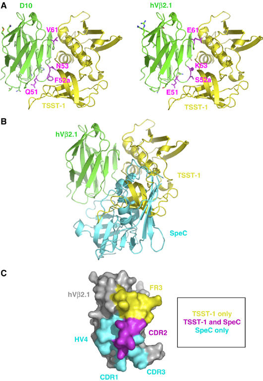
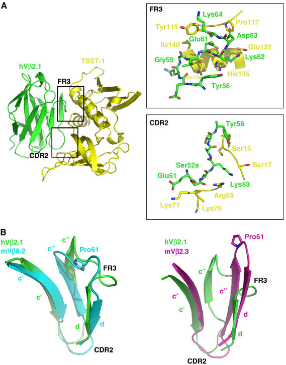
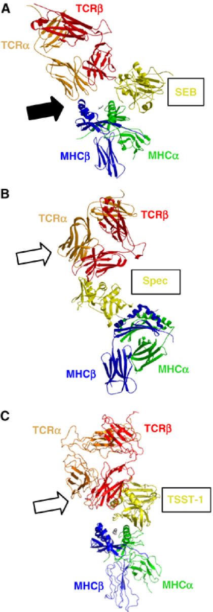
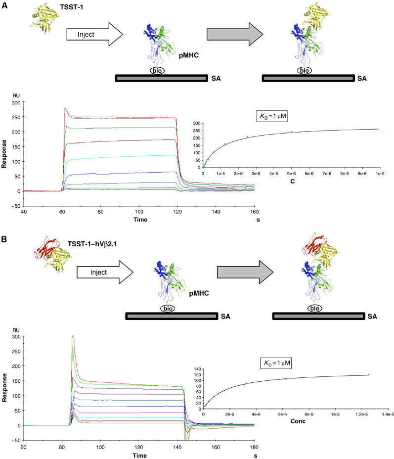
Superantigens (SAGs) bind simultaneously to major histocompatibility complex (MHC) and T-cell receptor (TCR) molecules, resulting in the massive release of inflammatory cytokines that can lead to toxic shock syndrome (TSS) and death. A major causative agent of TSS is toxic shock syndrome toxin-1 (TSST-1), which is unique relative to other bacterial SAGs owing to its structural divergence and its stringent TCR specificity. Here, we report the crystal structure of TSST-1 in complex with an affinity-matured variant of its wild-type TCR ligand, human T-cell receptor beta chain variable domain 2.1. From this structure and a model of the wild-type complex, we show that TSST-1 engages TCR ligands in a markedly different way than do other SAGs. We provide a structural basis for the high TCR specificity of TSST-1 and present a model of the TSST-1-dependent MHC-SAG-TCR T-cell signaling complex that is structurally and energetically unique relative to those formed by other SAGs. Our data also suggest that protein plasticity plays an exceptionally significant role in this affinity maturation process that results in more than a 3000-fold increase in affinity.


Figures

Figure 1
Figure 1
Distinct SAG-binding sites on a common ligand. (A) Crystal structure of the TSST-1–D10 complex (left panel) and a model of the wild-type TSST-1–hVβ2.1 complex (right panel). TSST-1 is in yellow; hVβ2.1 is in green. The side chains of the residues that are mutated in D10 relative to wild-type hVβ2.1 are shown. Those mutations that are energetically significant are in magenta; the remaining mutations are in green. (B) Superposition of the TSST-1–hVβ2.1 and SpeC–hVβ2.1 complexes. The hVβ2.1 molecule belonging to the SpeC–hVβ2.1 complex has been removed for clarity. Color coding is as in (A); SpeC is in cyan. (C) Molecular surface of hVβ2.1 buried uniquely by TSST-1 (yellow) or SpeC (cyan) and the shared portion of the epitope (magenta). The hVβ2.1 molecule in (C) has been rotated approximately 90° clockwise about the vertical axis of the page relative to its orientation in (A) and (B).
Figure 2
Figure 2
Structural features responsible for SAG–TCR specificity. (A) The TSST-1–hVβ2.1 complex with the CDR2 and FR3 hot regions demarcated. Upper inset, close-up of the molecular interactions in the FR3 hot region. Lower inset, close-up of the molecular interactions in the CDR2 hot region. Only residues that form contacts are drawn. Color coding is as in Figure 1A. (B) Superposition of the c′, c″ and d β-strands including the CDR2 and FR3 of hVβ2.1 with mVβ8.2 (left panel) and with mVβ2.3 (right panel). The side chains of Pro61 in both mVβ8.2 and mVβ2.3 are shown. Colors are as follows: hVβ2.1, green; mVβ8.2, cyan; mVβ2.3, magenta.
Figure 3
Figure 3
MHC–SAG–TCR T-cell signaling complexes. (A) The SEB-mediated T-cell signaling complex. The filled arrow indicates the direct MHCβ–TCRα interface. In this orientation, the APC would be situated below the complex; the T cell above. (B) The SpeC-mediated T-cell signaling complex. The unfilled arrow indicates that no direct MHC–TCR interaction occurs in this complex. (C) The TSST-1-mediated T-cell signaling complex. The unfilled arrow indicates that no direct MHC–TCR interaction occurs in this complex. For all panels, colors are as follows: SAG, yellow; TCRα, orange; TCRβ, red; MHCα, green; MHCβ, blue; peptide, gray.
Figure 4
Figure 4
No through-SAG cooperativity exhibited in the TSST-1-dependent T-cell signaling complex. (A) SPR sensorgram of the interaction between TSST-1 and immobilized pMHC. Nonlinear regression analysis of maximal responses versus concentration is shown to the right of the sensorgram. The experimental design is depicted in the box at the top of the panel. (B) SPR sensorgram of the interaction the TSST-1–D10 complex and immobilized pMHC. Nonlinear regression analysis of maximal responses versus concentration is shown to the right of the sensorgram. The experimental design is depicted in the box at the top of the panel. SA, streptavidin; bio, biotin.
Figure 5
Figure 5
Relative conformational flexibility of the hVβ2.1-binding site for TSST-1. (A) Structure of the affinity-matured hVβ2.1 variant D10 in the region encompassing the c′ β-strand, CDR2, c″ β-strand and FR3 in complex with TSST-1 (top panel), or of the same region of wild-type hVβ2.1 in complex with SpeC (middle panel) or pMHC (bottom panel). (B) Structure of mVβ8.2 in the region encompassing the c′ β-strand, CDR2, c″ β-strand and FR3 either uncomplexed (top panel), in complex with SEB (middle panel) or with pMHC (bottom panel). Residues involved in main-chain hydrogen-bonding interactions that noncovalently bind the c′ and c″ β-strands together are indicated in each panel.

References

    1. Andersen PS, Lavoie PM, Sekaly RP, Churchill H, Kranz DM, Schlievert PM, Karjalainen K, Mariuzza RA (1999) Role of the T cell receptor alpha chain in stabilizing TCR–superantigen–MHC class II complexes. Immunity 10: 473–483 - PubMed
    1. Andersen PS, Schuck P, Sundberg EJ, Geisler C, Karjalainen K, Mariuzza RA (2002) Quantifying the energetics of cooperativity in a ternary protein complex. Biochemistry 41: 5177–5184 - PubMed
    1. Bentley GA, Boulot G, Karjalainen K, Mariuzza RA (1995) Crystal structure of the beta chain of a T cell antigen receptor. Science 267: 1984–1987 - PubMed
    1. Bergdoll MS, Crass BA, Reiser RF, Robbins RN, Davis JP (1981) A new staphylococcal enterotoxin, enterotoxin F, associated with toxic-shock-syndrome Staphylococcus aureus isolates. Lancet 1: 1017–1021 - PubMed
    1. Brunger AT, Adams PD, Clore GM, DeLano WL, Gros P, Grosse-Kunstleve RW, Jiang JS, Kuszewski J, Nilges M, Pannu NS, Read RJ, Rice LM, Simonson T, Warren GL (1998) Crystallography and NMR system: a new software suite for macromolecular structure determination. Acta Crystallogr, D 54: 905–921 - PubMed

Publication types